6A17

Crystal structure of CYP90B1 in complex with brassinazole


Experimental Data Snapshot

  • Method: X-RAY DIFFRACTION
  • Resolution: 2.30 Å
  • R-Value Free: 0.275 
  • R-Value Work: 0.234 
  • R-Value Observed: 0.236 

wwPDB Validation   3D Report Full Report


Ligand Structure Quality Assessment 


This is version 1.2 of the entry. See complete history


Literature

Structural insights into a key step of brassinosteroid biosynthesis and its inhibition.

Fujiyama, K.Hino, T.Kanadani, M.Watanabe, B.Jae Lee, H.Mizutani, M.Nagano, S.

(2019) Nat Plants 5: 589-594

  • DOI: https://doi.org/10.1038/s41477-019-0436-6
  • Primary Citation of Related Structures:  
    6A15, 6A16, 6A17, 6A18

  • PubMed Abstract: 

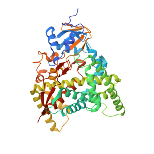
    Brassinosteroids (BRs) are essential plant steroid hormones that regulate plant growth and development 1 . The most potent BR, brassinolide, is produced by addition of many oxygen atoms to campesterol by several cytochrome P450 monooxygenases (CYPs). CYP90B1 (also known as DWF4) catalyses the 22(S)-hydroxylation of campesterol and is the first and rate-limiting enzyme at the branch point of the biosynthetic pathway from sterols to BRs 2 . Here we show the crystal structure of Arabidopsis thaliana CYP90B1 complexed with cholesterol as a substrate. The substrate-binding conformation explains the stereoselective introduction of a hydroxy group at the 22S position, facilitating hydrogen bonding of brassinolide with the BR receptor 3-5 . We also determined the crystal structures of CYP90B1 complexed with uniconazole 6,7 or brassinazole 8 , which inhibit BR biosynthesis. The two inhibitors are structurally similar; however, their binding conformations are unexpectedly different. The shape and volume of the active site pocket varies depending on which inhibitor or substrate is bound. These crystal structures of plant CYPs that function as membrane-anchored enzymes and exhibit structural plasticity can inform design of novel inhibitors targeting plant membrane-bound CYPs, including those involved in BR biosynthesis, which could then be used as plant growth regulators and agrochemicals.


  • Organizational Affiliation

    Department of Chemistry and Biotechnology, Graduate School of Engineering, Tottori University, Tottori, Japan.


Macromolecules
Find similar proteins by:  (by identity cutoff)  |  3D Structure
Entity ID: 1
MoleculeChains Sequence LengthOrganismDetailsImage
Cytochrome P450 90B1457Arabidopsis thalianaMutation(s): 1 
Gene Names: CYP90B1DWF4At3g50660T3A5.40
EC: 1.14
Membrane Entity: Yes 
UniProt
Find proteins for O64989 (Arabidopsis thaliana)
Explore O64989 
Go to UniProtKB:  O64989
Entity Groups  
Sequence Clusters30% Identity50% Identity70% Identity90% Identity95% Identity100% Identity
UniProt GroupO64989
Sequence Annotations
Expand
  • Reference Sequence
Small Molecules
Ligands 4 Unique
IDChains Name / Formula / InChI Key2D Diagram3D Interactions
HEM (Subject of Investigation/LOI)
Query on HEM

Download Ideal Coordinates CCD File 
C [auth A]PROTOPORPHYRIN IX CONTAINING FE
C34 H32 Fe N4 O4
KABFMIBPWCXCRK-RGGAHWMASA-L
9RL (Subject of Investigation/LOI)
Query on 9RL

Download Ideal Coordinates CCD File 
B [auth A](2R,3S)-4-(4-chlorophenyl)-2-phenyl-3-(1H-1,2,4-triazol-1-yl)butan-2-ol
C18 H18 Cl N3 O
YULDTPKHZNKFEY-ZWKOTPCHSA-N
GOL
Query on GOL

Download Ideal Coordinates CCD File 
D [auth A],
E [auth A]
GLYCEROL
C3 H8 O3
PEDCQBHIVMGVHV-UHFFFAOYSA-N
CL
Query on CL

Download Ideal Coordinates CCD File 
F [auth A],
G [auth A]
CHLORIDE ION
Cl
VEXZGXHMUGYJMC-UHFFFAOYSA-M
Binding Affinity Annotations 
IDSourceBinding Affinity
9RL Binding MOAD:  6A17 Ki: 1050 (nM) from 1 assay(s)
Experimental Data & Validation

Experimental Data

  • Method: X-RAY DIFFRACTION
  • Resolution: 2.30 Å
  • R-Value Free: 0.275 
  • R-Value Work: 0.234 
  • R-Value Observed: 0.236 
  • Space Group: P 61 2 2
Unit Cell:
Length ( Å )Angle ( ˚ )
a = 58.857α = 90
b = 58.857β = 90
c = 539.215γ = 120
Software Package:
Software NamePurpose
XDSdata reduction
XDSdata scaling
Aimlessdata scaling
PHASERphasing
Cootmodel building
PHENIXrefinement

Structure Validation

View Full Validation Report


Ligand Structure Quality Assessment 


Entry History & Funding Information

Deposition Data


Funding OrganizationLocationGrant Number
Japan Society for the Promotion of ScienceJapan17H05444

Revision History  (Full details and data files)

  • Version 1.0: 2019-06-12
    Type: Initial release
  • Version 1.1: 2019-07-10
    Changes: Data collection, Database references
  • Version 1.2: 2023-11-22
    Changes: Data collection, Database references, Refinement description