


Implications for tetraspanin-enriched microdomain assembly based on structures of CD9 with EWI-F.
Oosterheert, W., Xenaki, K.T., Neviani, V., Pos, W., Doulkeridou, S., Manshande, J., Pearce, N.M., Kroon-Batenburg, L.M., Lutz, M., van Bergen En Henegouwen, P.M., Gros, P.(2020) Life Sci Alliance 3
- PubMed: 32958604
- DOI: https://doi.org/10.26508/lsa.202000883
- Primary Citation of Related Structures:
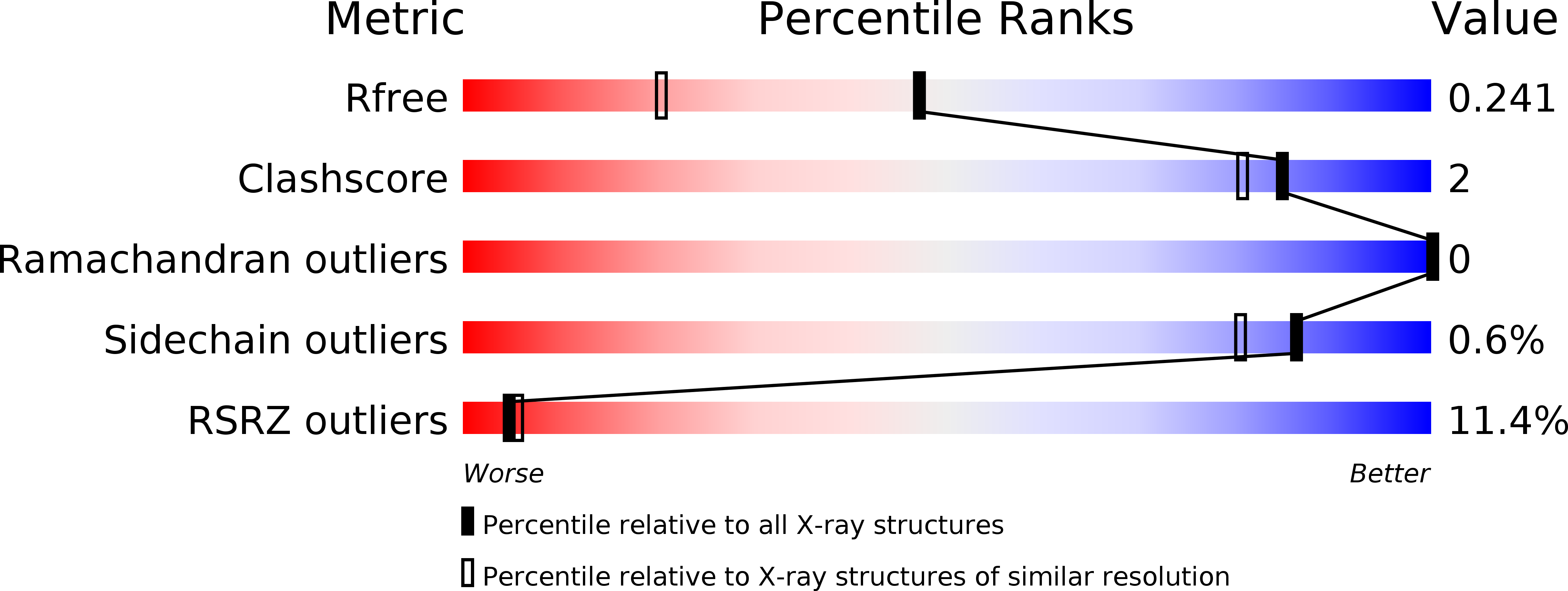
6RLR, 6Z1V, 6Z1Z, 6Z20 - PubMed Abstract:
Tetraspanins are eukaryotic membrane proteins that contribute to a variety of signaling processes by organizing partner-receptor molecules in the plasma membrane. How tetraspanins bind and cluster partner receptors into tetraspanin-enriched microdomains is unknown. Here, we present crystal structures of the large extracellular loop of CD9 bound to nanobodies 4C8 and 4E8 and, the cryo-EM structure of 4C8-bound CD9 in complex with its partner EWI-F. CD9-EWI-F displays a tetrameric arrangement with two central EWI-F molecules, dimerized through their ectodomains, and two CD9 molecules, one bound to each EWI-F transmembrane helix through CD9-helices h3 and h4. In the crystal structures, nanobodies 4C8 and 4E8 bind CD9 at loops C and D, which is in agreement with the 4C8 conformation in the CD9-EWI-F complex. The complex varies from nearly twofold symmetric (with the two CD9 copies nearly anti-parallel) to ca. 50° bent arrangements. This flexible arrangement of CD9-EWI-F with potential CD9 homo-dimerization at either end provides a "concatenation model" for forming short linear or circular assemblies, which may explain the occurrence of tetraspanin-enriched microdomains.
Organizational Affiliation:
Department of Chemistry, Crystal and Structural Chemistry, Bijvoet Centre for Biomolecular Research, Faculty of Science, Utrecht University, Utrecht, The Netherlands.


