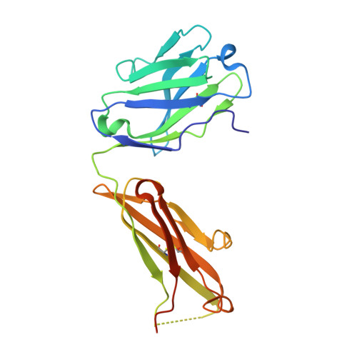
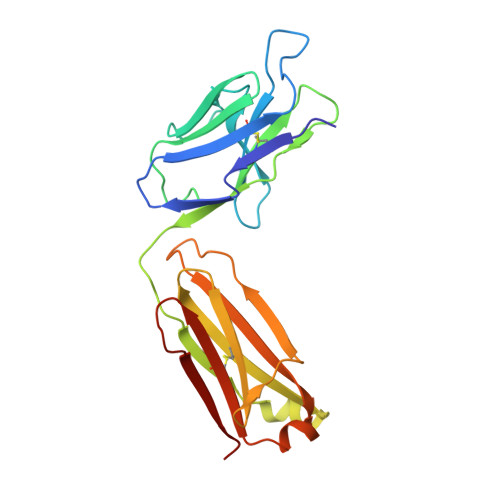
6Y54

Crystal structure of a Neisseria meningitidis serogroup A capsular oligosaccharide bound to a functional Fab


Experimental Data Snapshot

  • Method: X-RAY DIFFRACTION
  • Resolution: 2.67 Å
  • R-Value Free: 0.267 
  • R-Value Work: 0.210 
  • R-Value Observed: 0.213 

wwPDB Validation   3D Report Full Report


Ligand Structure Quality Assessment 


This is version 1.3 of the entry. See complete history


Literature

Structure of a protective epitope reveals the importance of acetylation of Neisseria meningitidis serogroup A capsular polysaccharide.

Henriques, P.Dello Iacono, L.Gimeno, A.Biolchi, A.Romano, M.R.Arda, A.Bernardes, G.J.L.Jimenez-Barbero, J.Berti, F.Rappuoli, R.Adamo, R.

(2020) Proc Natl Acad Sci U S A 117: 29795-29802

  • DOI: https://doi.org/10.1073/pnas.2011385117
  • Primary Citation of Related Structures:  
    6Y54

  • PubMed Abstract: 

    Meningococcal meningitis remains a substantial cause of mortality and morbidity worldwide. Until recently, countries in the African meningitis belt were susceptible to devastating outbreaks, largely attributed to serogroup A Neisseria meningitidis (MenA). Vaccination with glycoconjugates of MenA capsular polysaccharide led to an almost complete elimination of MenA clinical cases. To understand the molecular basis of vaccine-induced protection, we generated a panel of oligosaccharide fragments of different lengths and tested them with polyclonal and monoclonal antibodies by inhibition enzyme-linked immunosorbent assay, surface plasmon resonance, and competitive human serum bactericidal assay, which is a surrogate for protection. The epitope was shown to optimize between three and six repeating units and to be O -acetylated. The molecular interactions between a protective monoclonal antibody and a MenA capsular polysaccharide fragment were further elucidated at the atomic level by saturation transfer difference NMR spectroscopy and X-ray crystallography. The epitope consists of a trisaccharide anchored to the antibody via the O - and N -acetyl moieties through either H-bonding or CH-π interactions. In silico docking showed that 3- O -acetylation of the upstream residue is essential for antibody binding, while O -acetate could be equally accommodated at three and four positions of the other two residues. These results shed light on the mechanism of action of current MenA vaccines and provide a foundation for the rational design of improved therapies.


  • Organizational Affiliation

    Instituto de Medicina Molecular João Lobo Antunes, Faculdade de Medicina, Universidade de Lisboa, 1649-028 Lisboa, Portugal.


Macromolecules
Find similar proteins by:  (by identity cutoff)  |  3D Structure
Entity ID: 1
MoleculeChains Sequence LengthOrganismDetailsImage
Fab A1.1 H chainA [auth H],
C [auth M],
E [auth O]
226Mus musculusMutation(s): 0 
Entity Groups  
Sequence Clusters30% Identity50% Identity70% Identity90% Identity95% Identity100% Identity
Sequence Annotations
Expand
  • Reference Sequence
Find similar proteins by:  (by identity cutoff)  |  3D Structure
Entity ID: 2
MoleculeChains Sequence LengthOrganismDetailsImage
Fab A1.1 L chainB [auth L],
D [auth N],
F [auth P]
219Mus musculusMutation(s): 0 
Entity Groups  
Sequence Clusters30% Identity50% Identity70% Identity90% Identity95% Identity100% Identity
Sequence Annotations
Expand
  • Reference Sequence
Small Molecules
Ligands 5 Unique
IDChains Name / Formula / InChI Key2D Diagram3D Interactions
OOW
Query on OOW

Download Ideal Coordinates CCD File 
K [auth H],
U [auth M],
Y [auth O]
{[(2R,3S,4R,5S,6R)-5-acetamido-3-hydroxy-4-(2-oxopropyl)-6-(phosphonooxy)oxan-2-yl]methoxy}phosphonic acid
C10 H19 N O13 P2
WZRFFNAOXAJBLK-JDDHQFAOSA-N
OAB
Query on OAB

Download Ideal Coordinates CCD File 
AA [auth P],
O [auth L],
X [auth N]
{[(2R,3S,4R,5S,6S)-5-acetamido-3,6-dihydroxy-4-(2-oxopropyl)oxan-2-yl]methoxy}phosphonic acid
C10 H18 N O10 P
LMXWGHPJIXYEIE-QWIPYHCPSA-N
OA8
Query on OA8

Download Ideal Coordinates CCD File 
N [auth L],
W [auth N],
Z [auth P]
{[(2R,3S,4R,5S,6S)-3-(acetyloxy)-5-acetamido-4,6-dihydroxyoxan-2-yl]methoxy}phosphonic acid
C10 H18 N O10 P
WZFGOBLYIBEZRF-QWIPYHCPSA-N
IOD
Query on IOD

Download Ideal Coordinates CCD File 
G [auth H]
H
L
M [auth L]
P [auth M]
G [auth H],
H,
L,
M [auth L],
P [auth M],
Q [auth M],
R [auth M],
S [auth M],
V [auth N]
IODIDE ION
I
XMBWDFGMSWQBCA-UHFFFAOYSA-M
EDO
Query on EDO

Download Ideal Coordinates CCD File 
I [auth H],
J [auth H],
T [auth M]
1,2-ETHANEDIOL
C2 H6 O2
LYCAIKOWRPUZTN-UHFFFAOYSA-N
Experimental Data & Validation

Experimental Data

  • Method: X-RAY DIFFRACTION
  • Resolution: 2.67 Å
  • R-Value Free: 0.267 
  • R-Value Work: 0.210 
  • R-Value Observed: 0.213 
  • Space Group: P 1 21 1
Unit Cell:
Length ( Å )Angle ( ˚ )
a = 52.609α = 90
b = 153.205β = 91.527
c = 101.051γ = 90
Software Package:
Software NamePurpose
PHENIXrefinement
XDSdata reduction
Aimlessdata scaling
MOLREPphasing

Structure Validation

View Full Validation Report


Ligand Structure Quality Assessment 


Entry History & Funding Information

Deposition Data


Funding OrganizationLocationGrant Number
European CommissionItaly675671

Revision History  (Full details and data files)

  • Version 1.0: 2020-10-07
    Type: Initial release
  • Version 1.1: 2020-11-18
    Changes: Database references
  • Version 1.2: 2020-12-02
    Changes: Database references
  • Version 1.3: 2024-01-24
    Changes: Advisory, Data collection, Database references, Refinement description