6Y4S

Human kallikrein-related peptidase 7 (KLK7) in the unliganded state


Experimental Data Snapshot

  • Method: X-RAY DIFFRACTION
  • Resolution: 2.23 Å
  • R-Value Free: 0.242 
  • R-Value Work: 0.219 
  • R-Value Observed: 0.220 

wwPDB Validation   3D Report Full Report


This is version 1.2 of the entry. See complete history

Re-refinement Note

This entry reflects an alternative modeling of the original data in: 3BSQ


Literature

Structural Studies on the Inhibitory Binding Mode of Aromatic Coumarinic Esters to Human Kallikrein-Related Peptidase 7.

Hanke, S.Tindall, C.A.Pippel, J.Ulbricht, D.Pirotte, B.Reboud-Ravaux, M.Heiker, J.T.Strater, N.

(2020) J Med Chem 63: 5723-5733

  • DOI: https://doi.org/10.1021/acs.jmedchem.9b01806
  • Primary Citation of Related Structures:  
    6SHH, 6SHI, 6SJU, 6Y4S

  • PubMed Abstract: 

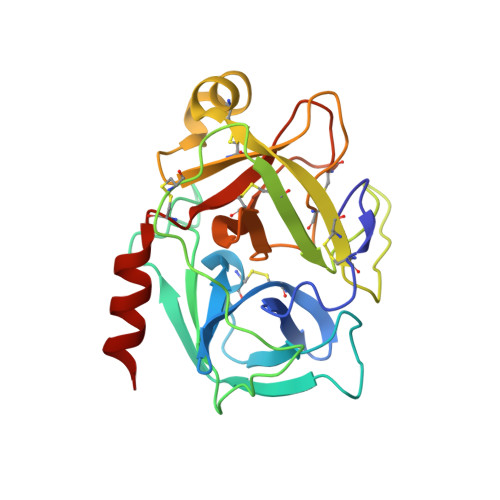
    The serine protease kallikrein-related peptidase 7 (KLK7) is a member of the human tissue kallikreins. Its dysregulation leads to pathophysiological inflammatory processes in the skin. Furthermore, it plays a role in several types of cancer. For the treatment of KLK7-associated diseases, coumarinic esters have been developed as small-molecule enzyme inhibitors. To characterize the inhibition mode of these inhibitors, we analyzed structures of the inhibited protease by X-ray crystallography. Electron density shows the inhibitors covalently attached to His57 of the catalytic triad. This confirms the irreversible character of the inhibition process. Upon inhibitor binding, His57 undergoes an outward rotation; thus, the catalytic triad of the protease is disrupted. Besides, the halophenyl moiety of the inhibitor was absent in the final enzyme-inhibitor complex due to the hydrolysis of the ester linkage. With these results, we analyze the structural basis of KLK7 inhibition by the covalent attachment of aromatic coumarinic esters.


  • Organizational Affiliation

    Institute of Bioanalytical Chemistry, Center for Biotechnology and Biomedicine, Leipzig University, Deutscher Platz 5, 04103 Leipzig, Germany.


Macromolecules
Find similar proteins by:  (by identity cutoff)  |  3D Structure
Entity ID: 1
MoleculeChains Sequence LengthOrganismDetailsImage
Kallikrein-7
A, B, C
224Homo sapiensMutation(s): 0 
Gene Names: KLK7PRSS6SCCE
EC: 3.4.21.117
UniProt & NIH Common Fund Data Resources
Find proteins for P49862 (Homo sapiens)
Explore P49862 
Go to UniProtKB:  P49862
PHAROS:  P49862
GTEx:  ENSG00000169035 
Entity Groups  
Sequence Clusters30% Identity50% Identity70% Identity90% Identity95% Identity100% Identity
UniProt GroupP49862
Sequence Annotations
Expand
  • Reference Sequence
Small Molecules
Ligands 2 Unique
IDChains Name / Formula / InChI Key2D Diagram3D Interactions
PGE
Query on PGE

Download Ideal Coordinates CCD File 
D [auth A]TRIETHYLENE GLYCOL
C6 H14 O4
ZIBGPFATKBEMQZ-UHFFFAOYSA-N
SO4
Query on SO4

Download Ideal Coordinates CCD File 
E [auth A]
F [auth A]
G [auth A]
H [auth A]
I [auth A]
E [auth A],
F [auth A],
G [auth A],
H [auth A],
I [auth A],
J [auth A],
K [auth B],
L [auth B],
M [auth B],
N [auth C],
O [auth C],
P [auth C],
Q [auth C],
R [auth C]
SULFATE ION
O4 S
QAOWNCQODCNURD-UHFFFAOYSA-L
Experimental Data & Validation

Experimental Data

  • Method: X-RAY DIFFRACTION
  • Resolution: 2.23 Å
  • R-Value Free: 0.242 
  • R-Value Work: 0.219 
  • R-Value Observed: 0.220 
  • Space Group: H 3 2
Unit Cell:
Length ( Å )Angle ( ˚ )
a = 113.15α = 90
b = 113.15β = 90
c = 326.14γ = 120
Software Package:
Software NamePurpose
BUSTERrefinement
XDSdata reduction
Aimlessdata scaling
PDB_EXTRACTdata extraction
REFMACphasing

Structure Validation

View Full Validation Report


Entry History 

Deposition Data

Revision History  (Full details and data files)

  • Version 1.0: 2020-05-20
    Type: Initial release
  • Version 1.1: 2020-06-24
    Changes: Database references
  • Version 1.2: 2024-01-24
    Changes: Data collection, Database references, Refinement description