Molecular and structural basis for Lewis glycan recognition by a cancer-targeting antibody.
Soliman, C., Guy, A.J., Chua, J.X., Vankemmelbeke, M., McIntosh, R.S., Eastwood, S., Truong, V.K., Elbourne, A., Spendlove, I., Durrant, L.G., Ramsland, P.A.(2020) Biochem J 477: 3219-3235
- PubMed: 32789497
- DOI: https://doi.org/10.1042/BCJ20200454
- Primary Citation of Related Structures:
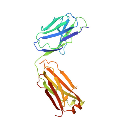
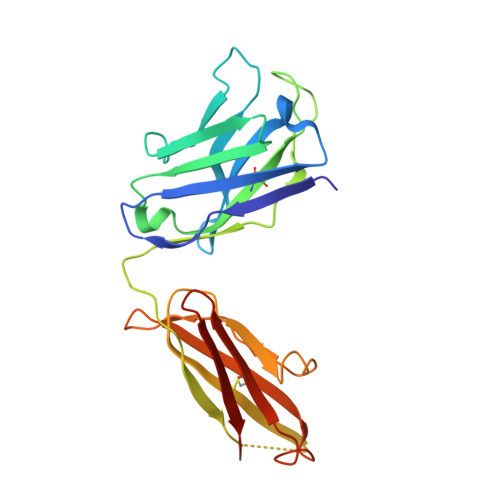
6X5E - PubMed Abstract:
Immunotherapy has been successful in treating many tumour types. The development of additional tumour-antigen binding monoclonal antibodies (mAbs) will help expand the range of immunotherapeutic targets. Lewis histo-blood group and related glycans are overexpressed on many carcinomas, including those of the colon, lung, breast, prostate and ovary, and can therefore be selectively targeted by mAbs. Here we examine the molecular and structural basis for recognition of extended Lea and Lex containing glycans by a chimeric mAb. Both the murine (FG88.2) IgG3 and a chimeric (ch88.2) IgG1 mAb variants showed reactivity to colorectal cancer cells leading to significantly reduced cell viability. We determined the X-ray structure of the unliganded ch88.2 fragment antigen-binding (Fab) containing two Fabs in the unit cell. A combination of molecular docking, glycan grafting and molecular dynamics simulations predicts two distinct subsites for recognition of Lea and Lex trisaccharides. While light chain residues were exclusively used for Lea binding, recognition of Lex involved both light and heavy chain residues. An extended groove is predicted to accommodate the Lea-Lex hexasaccharide with adjoining subsites for each trisaccharide. The molecular and structural details of the ch88.2 mAb presented here provide insight into its cross-reactivity for various Lea and Lex containing glycans. Furthermore, the predicted interactions with extended epitopes likely explains the selectivity of this antibody for targeting Lewis-positive tumours.
Organizational Affiliation:
School of Science, RMIT University, Melbourne, Victoria, Australia.