A Structural Systems Biology Approach to High-Risk CG23 Klebsiella pneumoniae.
Inniss, N.L., Kochan, T.J., Minasov, G., Wawrzak, Z., Chang, C., Tan, K., Shuvalova, L., Kiryukhina, O., Pshenychnyi, S., Wu, R., Dubrovska, I., Babnigg, G., Endres, M., Anderson, W.F., Hauser, A.R., Joachimiak, A., Satchell, K.J.F.(2023) Microbiol Resour Announc 12: e0101322-e0101322
- PubMed: 36695589
- DOI: https://doi.org/10.1128/mra.01013-22
- Primary Citation of Related Structures:
6DT3, 6DUX, 6DVV, 6DXN, 6E85, 6NAU, 6NBG, 6NDI, 6WN5, 6WN8, 6X1L, 7RJJ, 7TL5, 7TZP - PubMed Abstract:
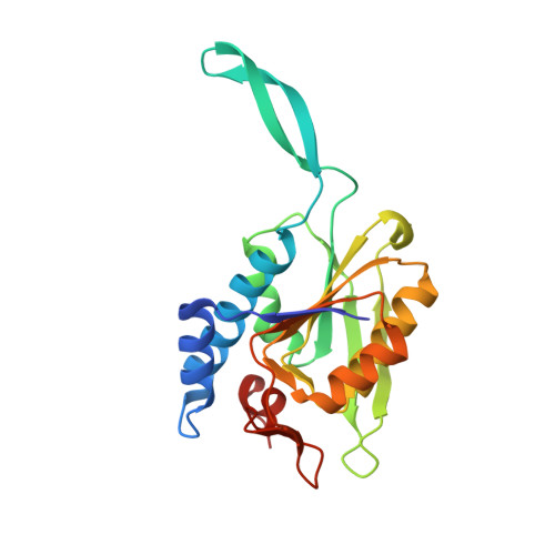
Klebsiella pneumoniae is a leading cause of antibiotic-resistant-associated deaths in the world. Here, we report the deposition of 14 structures of enzymes from both the core and accessory genomes of sequence type 23 (ST23) K1 hypervirulent K. pneumoniae.
Organizational Affiliation:
Department of Microbiology-Immunology, Feinberg School of Medicine, Northwestern University, Chicago, Illinois, USA.