


6WJ1
Crystal structure of Fab 54-4H03 bound to H1 influenza hemagglutinin
- PDB DOI: https://doi.org/10.2210/pdb6WJ1/pdb
- Classification: IMMUNE SYSTEM
- Organism(s): Influenza A virus, Homo sapiens
- Expression System: Trichoplusia ni, Cricetulus griseus
- Mutation(s): No
- Deposited: 2020-04-11 Released: 2020-07-01
- Funding Organization(s): National Institutes of Health/National Institute Of Allergy and Infectious Diseases (NIH/NIAID)
Experimental Data Snapshot
- Method: X-RAY DIFFRACTION
- Resolution: 3.50 Å
- R-Value Free: 0.232
- R-Value Work: 0.179
- R-Value Observed: 0.182

This is version 2.2 of the entry. See complete history.

Macromolecules
Find similar proteins by:
(by identity cutoff) | 3D Structure
Entity ID: 1 | |||||
|---|---|---|---|---|---|
| Molecule | Chains | Sequence Length | Organism | Details | Image |
| Hemagglutinin HA1 chain | A, B [auth C], C [auth E] | 330 | Influenza A virus | Mutation(s): 0 Gene Names: HA | 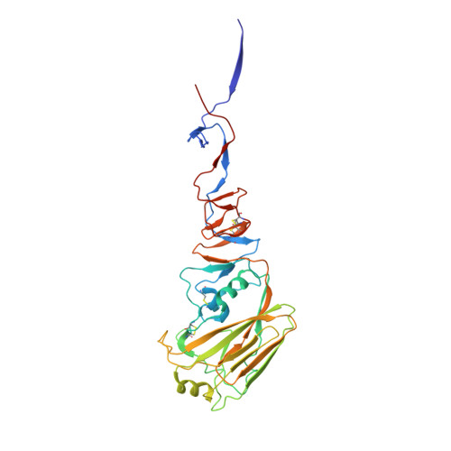 |
UniProt | |||||
Find proteins for C3W5S1 (Influenza A virus (strain swl A/California/04/2009 H1N1)) Explore C3W5S1 Go to UniProtKB: C3W5S1 | |||||
Entity Groups | |||||
| Sequence Clusters | 30% Identity50% Identity70% Identity90% Identity95% Identity100% Identity | ||||
| UniProt Group | C3W5S1 | ||||
Sequence AnnotationsExpand | |||||
| |||||
Find similar proteins by:
(by identity cutoff) | 3D Structure
Entity ID: 2 | |||||
|---|---|---|---|---|---|
| Molecule | Chains | Sequence Length | Organism | Details | Image |
| Hemagglutinin HA2 chain | D [auth B], E [auth D], F | 175 | Influenza A virus | Mutation(s): 0 Gene Names: HA | 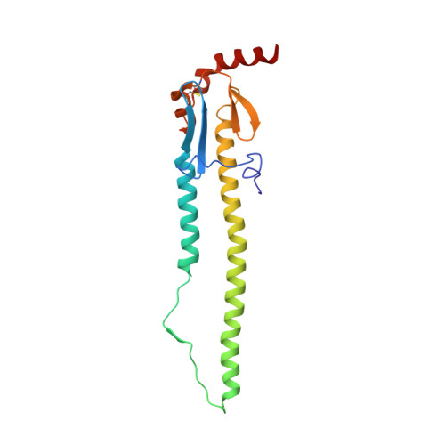 |
UniProt | |||||
Find proteins for C3W5S1 (Influenza A virus (strain swl A/California/04/2009 H1N1)) Explore C3W5S1 Go to UniProtKB: C3W5S1 | |||||
Entity Groups | |||||
| Sequence Clusters | 30% Identity50% Identity70% Identity90% Identity95% Identity100% Identity | ||||
| UniProt Group | C3W5S1 | ||||
Sequence AnnotationsExpand | |||||
| |||||
Find similar proteins by:
(by identity cutoff) | 3D Structure
Entity ID: 3 | |||||
|---|---|---|---|---|---|
| Molecule | Chains | Sequence Length | Organism | Details | Image |
| Fab 54-4H03 heavy chain | G [auth H], I [auth J], K [auth G] | 230 | Homo sapiens | Mutation(s): 0 | 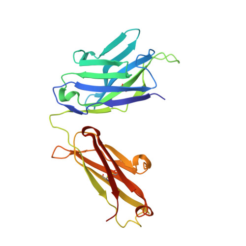 |
Entity Groups | |||||
| Sequence Clusters | 30% Identity50% Identity70% Identity90% Identity95% Identity100% Identity | ||||
Sequence AnnotationsExpand | |||||
| |||||
Find similar proteins by:
(by identity cutoff) | 3D Structure
Entity ID: 4 | |||||
|---|---|---|---|---|---|
| Molecule | Chains | Sequence Length | Organism | Details | Image |
| Fab 54-4H03 light chain | H [auth L], J [auth K], L [auth I] | 216 | Homo sapiens | Mutation(s): 0 | 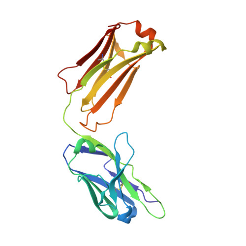 |
Entity Groups | |||||
| Sequence Clusters | 30% Identity50% Identity70% Identity90% Identity95% Identity100% Identity | ||||
Sequence AnnotationsExpand | |||||
| |||||
Oligosaccharides
Entity ID: 5 | |||||
|---|---|---|---|---|---|
| Molecule | Chains | Length | 2D Diagram | Glycosylation | 3D Interactions |
| 2-acetamido-2-deoxy-beta-D-glucopyranose-(1-4)-2-acetamido-2-deoxy-beta-D-glucopyranose | M, O, T | 2 |  | N-Glycosylation | |
Glycosylation Resources | |||||
GlyTouCan: G42666HT GlyCosmos: G42666HT GlyGen: G42666HT | |||||
Entity ID: 6 | |||||
|---|---|---|---|---|---|
| Molecule | Chains | Length | 2D Diagram | Glycosylation | 3D Interactions |
| alpha-D-mannopyranose-(1-3)-[alpha-D-mannopyranose-(1-6)]beta-D-mannopyranose-(1-4)-2-acetamido-2-deoxy-beta-D-glucopyranose-(1-4)-2-acetamido-2-deoxy-beta-D-glucopyranose | N | 5 |  | N-Glycosylation | |
Glycosylation Resources | |||||
GlyTouCan: G22768VO GlyCosmos: G22768VO GlyGen: G22768VO | |||||
Entity ID: 7 | |||||
|---|---|---|---|---|---|
| Molecule | Chains | Length | 2D Diagram | Glycosylation | 3D Interactions |
| alpha-D-mannopyranose-(1-6)-beta-D-mannopyranose-(1-4)-2-acetamido-2-deoxy-beta-D-glucopyranose-(1-4)-2-acetamido-2-deoxy-beta-D-glucopyranose | P | 4 |  | N-Glycosylation | |
Glycosylation Resources | |||||
GlyTouCan: G22573RC GlyCosmos: G22573RC GlyGen: G22573RC | |||||
Entity ID: 8 | |||||
|---|---|---|---|---|---|
| Molecule | Chains | Length | 2D Diagram | Glycosylation | 3D Interactions |
| alpha-L-fucopyranose-(1-6)-2-acetamido-2-deoxy-beta-D-glucopyranose | Q | 2 |  | N-Glycosylation | |
Glycosylation Resources | |||||
GlyTouCan: G86851RC GlyCosmos: G86851RC GlyGen: G86851RC | |||||
Entity ID: 9 | |||||
|---|---|---|---|---|---|
| Molecule | Chains | Length | 2D Diagram | Glycosylation | 3D Interactions |
| alpha-D-mannopyranose-(1-3)-beta-D-mannopyranose-(1-4)-2-acetamido-2-deoxy-beta-D-glucopyranose-(1-4)-2-acetamido-2-deoxy-beta-D-glucopyranose | R | 4 |  | N-Glycosylation | |
Glycosylation Resources | |||||
GlyTouCan: G81315DD GlyCosmos: G81315DD GlyGen: G81315DD | |||||

Small Molecules
| Ligands 1 Unique | |||||
|---|---|---|---|---|---|
| ID | Chains | Name / Formula / InChI Key | 2D Diagram | 3D Interactions | |
| NAG Query on NAG | AA [auth E] BA [auth E] CA [auth B] DA [auth D] U [auth A] | 2-acetamido-2-deoxy-beta-D-glucopyranose C8 H15 N O6 OVRNDRQMDRJTHS-FMDGEEDCSA-N |  | ||
Experimental Data & Validation
Experimental Data
- Method: X-RAY DIFFRACTION
- Resolution: 3.50 Å
- R-Value Free: 0.232
- R-Value Work: 0.179
- R-Value Observed: 0.182
- Space Group: C 2 2 21
Unit Cell:
| Length ( Å ) | Angle ( ˚ ) |
|---|---|
| a = 231.538 | α = 90 |
| b = 259.211 | β = 90 |
| c = 165.113 | γ = 90 |
| Software Name | Purpose |
|---|---|
| PHENIX | refinement |
| HKL-2000 | data reduction |
| PDB_EXTRACT | data extraction |
| PHASER | phasing |
Entry History & Funding Information
Deposition Data
- Released Date: 2020-07-01 Deposition Author(s): Wu, N.C., Wilson, I.A.
| Funding Organization | Location | Grant Number |
|---|---|---|
| National Institutes of Health/National Institute Of Allergy and Infectious Diseases (NIH/NIAID) | United States | R56 AI127371 |
| National Institutes of Health/National Institute Of Allergy and Infectious Diseases (NIH/NIAID) | United States | K99 AI139445 |
Revision History (Full details and data files)
- Version 1.0: 2020-07-01
Type: Initial release - Version 1.1: 2020-07-15
Changes: Advisory, Database references, Derived calculations - Version 2.0: 2020-07-29
Type: Remediation
Reason: Carbohydrate remediation
Changes: Advisory, Atomic model, Data collection, Derived calculations, Structure summary - Version 2.1: 2020-09-23
Changes: Database references, Structure summary - Version 2.2: 2023-10-18
Changes: Data collection, Database references, Refinement description
