6W7S

Ketoreductase from module 1 of the 6-deoxyerythronolide B synthase (KR1) in complex with antibody fragment (Fab) 2G10


Experimental Data Snapshot

  • Method: X-RAY DIFFRACTION
  • Resolution: 2.25 Å
  • R-Value Free: 0.232 
  • R-Value Work: 0.179 
  • R-Value Observed: 0.182 

wwPDB Validation   3D Report Full Report


This is version 1.2 of the entry. See complete history


Literature

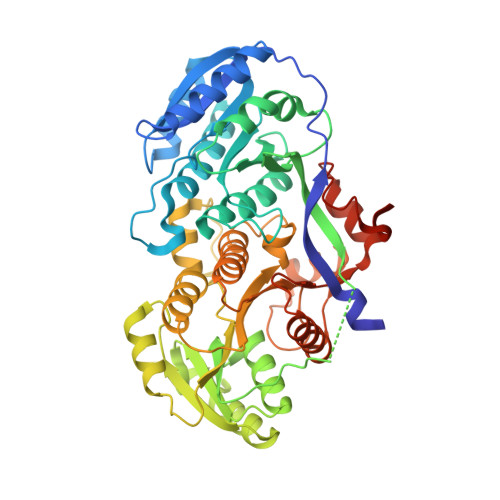
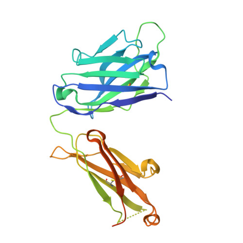
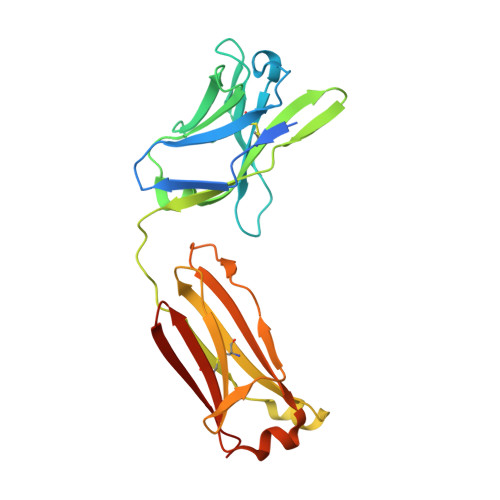
Antibody Probes of Module 1 of the 6-Deoxyerythronolide B Synthase Reveal an Extended Conformation During Ketoreduction.

Cogan, D.P.Li, X.Sevillano, N.Mathews, I.I.Matsui, T.Craik, C.S.Khosla, C.

(2020) J Am Chem Soc 142: 14933-14939

  • DOI: https://doi.org/10.1021/jacs.0c05133
  • Primary Citation of Related Structures:  
    6W7S, 6WH9

  • PubMed Abstract: 

    The 6-deoxyerythronolide B synthase (DEBS) is a prototypical assembly line polyketide synthase (PKS) that synthesizes the macrocyclic core of the antibiotic erythromycin. Each of its six multidomain modules presumably sample distinct conformations, as biosynthetic intermediates tethered to their acyl carrier proteins interact with multiple active sites during the courses of their catalytic cycles. The spatiotemporal details underlying these protein dynamics remain elusive. Here, we investigate one aspect of this conformational flexibility using two domain-specific monoclonal antibody fragments (F ab s) isolated from a very large naïve human antibody library. Both F ab s, designated 1D10 and 2G10, were bound specifically and with high affinity to the ketoreductase domain of DEBS module 1 (KR1). Comparative kinetic analysis of stand-alone KR1 as well as a truncated bimodular derivative of DEBS revealed that 1D10 inhibited KR1 activity whereas 2G10 did not. Co-crystal structures of each KR1-F ab complex provided a mechanistic rationale for this difference. A hybrid PKS module harboring KR1 was engineered, whose individual catalytic domains have been crystallographically characterized at high resolution. Size exclusion chromatography coupled to small-angle X-ray scattering (SEC-SAXS) of this hybrid module bound to 1D10 provided further support for the catalytic relevance of the "extended" model of a PKS module. Our findings reinforce the power of monoclonal antibodies as tools to interrogate structure-function relationships of assembly line PKSs.


  • Organizational Affiliation

    Department of Pharmaceutical Chemistry, University of California San Francisco, San Francisco, California 94158, United States.


Macromolecules
Find similar proteins by:  (by identity cutoff)  |  3D Structure
Entity ID: 1
MoleculeChains Sequence LengthOrganismDetailsImage
EryAI485Saccharopolyspora erythraeaMutation(s): 0 
Gene Names: eryAI
UniProt
Find proteins for Q03131 (Saccharopolyspora erythraea)
Explore Q03131 
Go to UniProtKB:  Q03131
Entity Groups  
Sequence Clusters30% Identity50% Identity70% Identity90% Identity95% Identity100% Identity
UniProt GroupQ03131
Sequence Annotations
Expand
  • Reference Sequence
Find similar proteins by:  (by identity cutoff)  |  3D Structure
Entity ID: 2
MoleculeChains Sequence LengthOrganismDetailsImage
2G10 (Fab heavy chain)B [auth H]255Homo sapiensMutation(s): 0 
Entity Groups  
Sequence Clusters30% Identity50% Identity70% Identity90% Identity95% Identity100% Identity
Sequence Annotations
Expand
  • Reference Sequence
Find similar proteins by:  (by identity cutoff)  |  3D Structure
Entity ID: 3
MoleculeChains Sequence LengthOrganismDetailsImage
2G10 (Fab light chain)C [auth L]236Homo sapiensMutation(s): 0 
Entity Groups  
Sequence Clusters30% Identity50% Identity70% Identity90% Identity95% Identity100% Identity
Sequence Annotations
Expand
  • Reference Sequence
Small Molecules
Ligands 2 Unique
IDChains Name / Formula / InChI Key2D Diagram3D Interactions
MES
Query on MES

Download Ideal Coordinates CCD File 
D [auth A]2-(N-MORPHOLINO)-ETHANESULFONIC ACID
C6 H13 N O4 S
SXGZJKUKBWWHRA-UHFFFAOYSA-N
CL
Query on CL

Download Ideal Coordinates CCD File 
E [auth L]CHLORIDE ION
Cl
VEXZGXHMUGYJMC-UHFFFAOYSA-M
Experimental Data & Validation

Experimental Data

  • Method: X-RAY DIFFRACTION
  • Resolution: 2.25 Å
  • R-Value Free: 0.232 
  • R-Value Work: 0.179 
  • R-Value Observed: 0.182 
  • Space Group: C 2 2 21
Unit Cell:
Length ( Å )Angle ( ˚ )
a = 80.148α = 90
b = 129.532β = 90
c = 208.818γ = 90
Software Package:
Software NamePurpose
PHENIXrefinement
XDSdata reduction
Aimlessdata scaling
PDB_EXTRACTdata extraction
PHASERphasing

Structure Validation

View Full Validation Report


Entry History & Funding Information

Deposition Data


Funding OrganizationLocationGrant Number
National Institutes of Health/National Institute of General Medical Sciences (NIH/NIGMS)United StatesGM087934

Revision History  (Full details and data files)

  • Version 1.0: 2020-09-09
    Type: Initial release
  • Version 1.1: 2020-09-16
    Changes: Database references
  • Version 1.2: 2023-10-18
    Changes: Data collection, Database references, Refinement description