6W03

Crystal Structure of HIV-1 BG505 DS-SOSIP.3mut Prefusion Env Trimer in Complex with Human Antibodies 3H109L and 35O22 at 3.3 Angstrom


Experimental Data Snapshot

  • Method: X-RAY DIFFRACTION
  • Resolution: 2.40 Å
  • R-Value Free: 0.273 
  • R-Value Work: 0.230 
  • R-Value Observed: 0.233 

wwPDB Validation   3D Report Full Report

Currently 6W03 does not have a validation slider image.


This is version 2.2 of the entry. See complete history


Literature

Development of a 3Mut-Apex-Stabilized Envelope Trimer That Expands HIV-1 Neutralization Breadth When Used To Boost Fusion Peptide-Directed Vaccine-Elicited Responses.

Chuang, G.Y.Lai, Y.T.Boyington, J.C.Cheng, C.Geng, H.Narpala, S.Rawi, R.Schmidt, S.D.Tsybovsky, Y.Verardi, R.Xu, K.Yang, Y.Zhang, B.Chambers, M.Changela, A.Corrigan, A.R.Kong, R.Olia, A.S.Ou, L.Sarfo, E.K.Wang, S.Wu, W.Doria-Rose, N.A.McDermott, A.B.Mascola, J.R.Kwong, P.D.

(2020) J Virol 94

  • DOI: https://doi.org/10.1128/JVI.00074-20
  • Primary Citation of Related Structures:  
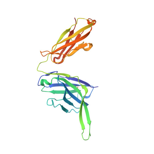
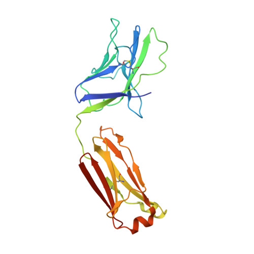
    6VZI, 6W03

  • PubMed Abstract: 

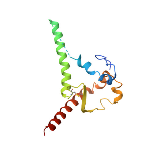
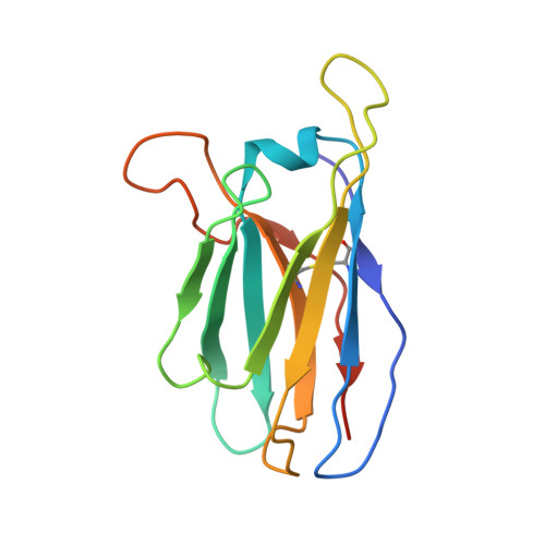
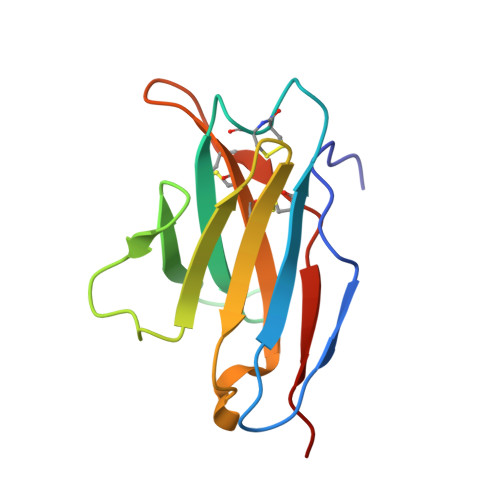
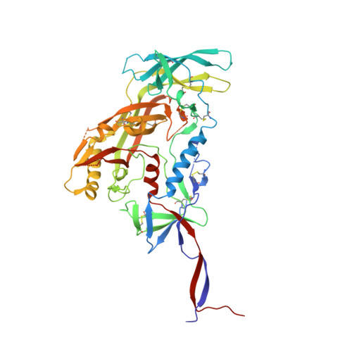
    HIV-1 envelope (Env) trimers, stabilized in a prefusion-closed conformation, can elicit humoral responses capable of neutralizing HIV-1 strains closely matched in sequence to the immunizing strain. One strategy to increase elicited neutralization breadth involves vaccine priming of immune responses against a target site of vulnerability, followed by vaccine boosting of these responses with prefusion-closed Env trimers. This strategy has succeeded at the fusion peptide (FP) site of vulnerability in eliciting cross-clade neutralizing responses in standard vaccine-test animals. However, the breadth and potency of the elicited responses have been less than optimal. Here, we identify three mutations (3mut), Met302, Leu320, and Pro329, that stabilize the apex of the Env trimer in a prefusion-closed conformation and show antigenically, structurally, and immunogenically that combining 3mut with other approaches (e.g., repair and stabilize and glycine-helix breaking) yields well-behaved clade C-Env trimers capable of boosting the breadth of FP-directed responses. Crystal structures of these trimers confirmed prefusion-closed apexes stabilized by hydrophobic patches contributed by Met302 and Leu320, with Pro329 assuming canonically restricted dihedral angles. We substituted the N-terminal eight residues of FP (FP8, residues 512 to 519) of these trimers with the second most prevalent FP8 sequence (FP8v2, AVGLGAVF) and observed a 3mut-stabilized consensus clade C-Env trimer with FP8v2 to boost the breadth elicited in guinea pigs of FP-directed responses induced by immunogens containing the most prevalent FP8 sequence (FP8v1, AVGIGAVF). Overall, 3mut can stabilize the Env trimer apex, and the resultant apex-stabilized Env trimers can be used to expand the neutralization breadth elicited against the FP site of vulnerability. IMPORTANCE A major hurdle to the development of an effective HIV-1 vaccine is the elicitation of serum responses capable of neutralizing circulating strains of HIV, which are extraordinarily diverse in sequence and often highly neutralization resistant. Recently, we showed how sera with 20 to 30% neutralization breadth could, nevertheless, be elicited in standard vaccine test animals by priming with the most prevalent N-terminal 8 residues of the HIV-1 fusion peptide (FP8), followed by boosting with a stabilized BG505-envelope (Env) trimer. Here, we show that subsequent boosting with a 3mut-apex-stabilized consensus C-Env trimer, modified to have the second most prevalent FP8 sequence, elicits higher neutralization breadth than that induced by continued boosting with the stabilized BG505-Env trimer. With increased neutralizing breadth elicited by boosting with a heterologous trimer containing the second most prevalent FP8 sequence, the fusion peptide-directed immune-focusing approach moves a step closer toward realizing an effective HIV-1 vaccine regimen.


  • Organizational Affiliation

    Vaccine Research Center, National Institute of Allergy and Infectious Diseases, National Institutes of Health, Bethesda, Maryland, USA.


Macromolecules
Find similar proteins by:  (by identity cutoff)  |  3D Structure
Entity ID: 1
MoleculeChains Sequence LengthOrganismDetailsImage
Envelope glycoprotein gp41A [auth B]153Human immunodeficiency virus 1Mutation(s): 2 
Gene Names: env
UniProt
Find proteins for Q2N0S6 (Human immunodeficiency virus 1)
Explore Q2N0S6 
Go to UniProtKB:  Q2N0S6
Entity Groups  
Sequence Clusters30% Identity50% Identity70% Identity90% Identity95% Identity100% Identity
UniProt GroupQ2N0S6
Sequence Annotations
Expand
  • Reference Sequence
Find similar proteins by:  (by identity cutoff)  |  3D Structure
Entity ID: 2
MoleculeChains Sequence LengthOrganismDetailsImage
35O22 scFv heavy chainB [auth D]134Homo sapiensMutation(s): 0 
Entity Groups  
Sequence Clusters30% Identity50% Identity70% Identity90% Identity95% Identity100% Identity
Sequence Annotations
Expand
  • Reference Sequence
Find similar proteins by:  (by identity cutoff)  |  3D Structure
Entity ID: 3
MoleculeChains Sequence LengthOrganismDetailsImage
35O22 scFv light chainC [auth E]114Homo sapiensMutation(s): 0 
Entity Groups  
Sequence Clusters30% Identity50% Identity70% Identity90% Identity95% Identity100% Identity
Sequence Annotations
Expand
  • Reference Sequence
Find similar proteins by:  (by identity cutoff)  |  3D Structure
Entity ID: 4
MoleculeChains Sequence LengthOrganismDetailsImage
Envelope glycoprotein gp160D [auth G]481Human immunodeficiency virus 1Mutation(s): 8 
Gene Names: env
UniProt
Find proteins for Q2N0S6 (Human immunodeficiency virus 1)
Explore Q2N0S6 
Go to UniProtKB:  Q2N0S6
Entity Groups  
Sequence Clusters30% Identity50% Identity70% Identity90% Identity95% Identity100% Identity
UniProt GroupQ2N0S6
Sequence Annotations
Expand
  • Reference Sequence
Find similar proteins by:  (by identity cutoff)  |  3D Structure
Entity ID: 5
MoleculeChains Sequence LengthOrganismDetailsImage
3H109L Fab heavy chainE [auth H]244Homo sapiensMutation(s): 0 
Entity Groups  
Sequence Clusters30% Identity50% Identity70% Identity90% Identity95% Identity100% Identity
Sequence Annotations
Expand
  • Reference Sequence
Find similar proteins by:  (by identity cutoff)  |  3D Structure
Entity ID: 6
MoleculeChains Sequence LengthOrganismDetailsImage
3H109L Fab light chainF [auth L]217Homo sapiensMutation(s): 0 
Entity Groups  
Sequence Clusters30% Identity50% Identity70% Identity90% Identity95% Identity100% Identity
Sequence Annotations
Expand
  • Reference Sequence
Oligosaccharides

Help

Entity ID: 7
MoleculeChains Length2D Diagram Glycosylation3D Interactions
alpha-D-mannopyranose-(1-6)-beta-D-mannopyranose-(1-4)-2-acetamido-2-deoxy-beta-D-glucopyranose-(1-4)-2-acetamido-2-deoxy-beta-D-glucopyranoseG [auth A]4N-Glycosylation
Glycosylation Resources
GlyTouCan:  G22573RC
GlyCosmos:  G22573RC
GlyGen:  G22573RC
Entity ID: 8
MoleculeChains Length2D Diagram Glycosylation3D Interactions
2-acetamido-2-deoxy-beta-D-glucopyranose-(1-4)-2-acetamido-2-deoxy-beta-D-glucopyranoseH [auth C],
I [auth F],
J [auth I],
K [auth J],
L [auth K]
2N-Glycosylation
Glycosylation Resources
GlyTouCan:  G42666HT
GlyCosmos:  G42666HT
GlyGen:  G42666HT
Entity ID: 9
MoleculeChains Length2D Diagram Glycosylation3D Interactions
alpha-D-mannopyranose-(1-3)-[alpha-D-mannopyranose-(1-6)]beta-D-mannopyranose-(1-4)-2-acetamido-2-deoxy-beta-D-glucopyranose-(1-4)-2-acetamido-2-deoxy-beta-D-glucopyranose
M
5N-Glycosylation
Glycosylation Resources
GlyTouCan:  G22768VO
GlyCosmos:  G22768VO
GlyGen:  G22768VO
Entity ID: 10
MoleculeChains Length2D Diagram Glycosylation3D Interactions
alpha-D-mannopyranose-(1-3)-beta-D-mannopyranose-(1-4)-2-acetamido-2-deoxy-beta-D-glucopyranose-(1-4)-2-acetamido-2-deoxy-beta-D-glucopyranose
N
4N-Glycosylation
Glycosylation Resources
GlyTouCan:  G81315DD
GlyCosmos:  G81315DD
GlyGen:  G81315DD
Entity ID: 11
MoleculeChains Length2D Diagram Glycosylation3D Interactions
alpha-D-mannopyranose-(1-3)-alpha-D-mannopyranose-(1-6)-[alpha-D-mannopyranose-(1-3)]beta-D-mannopyranose-(1-4)-2-acetamido-2-deoxy-beta-D-glucopyranose-(1-4)-2-acetamido-2-deoxy-beta-D-glucopyranose
O
6N-Glycosylation
Glycosylation Resources
GlyTouCan:  G09724ZC
GlyCosmos:  G09724ZC
GlyGen:  G09724ZC
Entity ID: 12
MoleculeChains Length2D Diagram Glycosylation3D Interactions
alpha-D-mannopyranose-(1-2)-alpha-D-mannopyranose-(1-2)-alpha-D-mannopyranose-(1-3)-[alpha-D-mannopyranose-(1-2)-alpha-D-mannopyranose-(1-6)-[alpha-D-mannopyranose-(1-3)]alpha-D-mannopyranose-(1-6)]beta-D-mannopyranose-(1-4)-2-acetamido-2-deoxy-beta-D-glucopyranose-(1-4)-2-acetamido-2-deoxy-beta-D-glucopyranose
P
10N-Glycosylation
Glycosylation Resources
GlyTouCan:  G40702WU
GlyCosmos:  G40702WU
GlyGen:  G40702WU
Experimental Data & Validation

Experimental Data

  • Method: X-RAY DIFFRACTION
  • Resolution: 2.40 Å
  • R-Value Free: 0.273 
  • R-Value Work: 0.230 
  • R-Value Observed: 0.233 
  • Space Group: P 63
Unit Cell:
Length ( Å )Angle ( ˚ )
a = 131.39α = 90
b = 131.39β = 90
c = 315.34γ = 120
Software Package:
Software NamePurpose
PHENIXrefinement
HKL-2000data scaling
PDB_EXTRACTdata extraction
HKL-2000data reduction
PHASERphasing

Structure Validation

View Full Validation Report

Currently 6W03 does not have a validation slider image.


Entry History 

Deposition Data

Revision History  (Full details and data files)

  • Version 1.0: 2020-04-15
    Type: Initial release
  • Version 2.0: 2020-07-29
    Type: Remediation
    Reason: Carbohydrate remediation
    Changes: Advisory, Atomic model, Data collection, Derived calculations, Structure summary
  • Version 2.1: 2021-10-27
    Changes: Database references, Structure summary
  • Version 2.2: 2023-10-11
    Changes: Data collection, Refinement description