


6VZZ
Crystal structure of glucokinase from Balamuthia mandrillaris in complex with glucose
- PDB DOI: https://doi.org/10.2210/pdb6VZZ/pdb
- Classification: TRANSFERASE
- Organism(s): Balamuthia mandrillaris
- Expression System: Escherichia coli BL21(DE3)
- Mutation(s): No
- Deposited: 2020-02-28 Released: 2020-03-25
Experimental Data Snapshot
- Method: X-RAY DIFFRACTION
- Resolution: 2.65 Å
- R-Value Free: 0.204
- R-Value Work: 0.174
- R-Value Observed: 0.177
wwPDB Validation 3D Report Full Report
Currently 6VZZ does not have a validation slider image.
This is version 1.3 of the entry. See complete history.

Macromolecules
Find similar proteins by:
(by identity cutoff) | 3D Structure
Entity ID: 1 | |||||
|---|---|---|---|---|---|
| Molecule | Chains | Sequence Length | Organism | Details | Image |
| BamaA.19900.a | 391 | Balamuthia mandrillaris | Mutation(s): 0 | 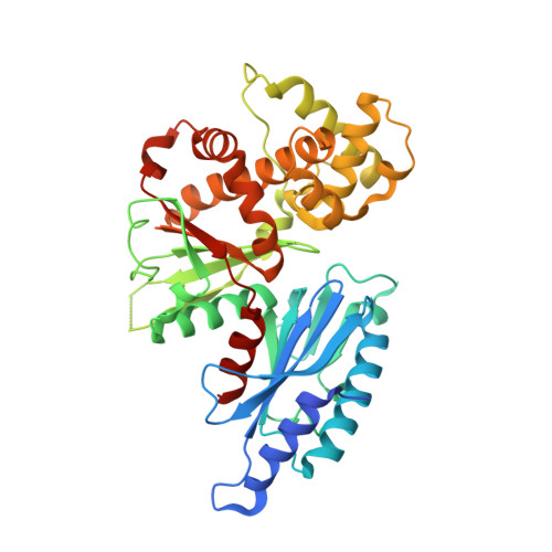 | |
Entity Groups | |||||
| Sequence Clusters | 30% Identity50% Identity70% Identity90% Identity95% Identity100% Identity | ||||
Sequence AnnotationsExpand | |||||
| |||||
Small Molecules
| Ligands 3 Unique | |||||
|---|---|---|---|---|---|
| ID | Chains | Name / Formula / InChI Key | 2D Diagram | 3D Interactions | |
| BGC (Subject of Investigation/LOI) Query on BGC | B [auth A] | beta-D-glucopyranose C6 H12 O6 WQZGKKKJIJFFOK-VFUOTHLCSA-N |  | ||
| EDO Query on EDO | F [auth A] | 1,2-ETHANEDIOL C2 H6 O2 LYCAIKOWRPUZTN-UHFFFAOYSA-N |  | ||
| CA Query on CA | C [auth A], D [auth A], E [auth A] | CALCIUM ION Ca BHPQYMZQTOCNFJ-UHFFFAOYSA-N |  | ||
Experimental Data & Validation
Experimental Data
- Method: X-RAY DIFFRACTION
- Resolution: 2.65 Å
- R-Value Free: 0.204
- R-Value Work: 0.174
- R-Value Observed: 0.177
- Space Group: P 41 3 2
- Diffraction Data: https://doi.org/10.18430/m36vzz
Unit Cell:
| Length ( Å ) | Angle ( ˚ ) |
|---|---|
| a = 157.3 | α = 90 |
| b = 157.3 | β = 90 |
| c = 157.3 | γ = 90 |
| Software Name | Purpose |
|---|---|
| PHENIX | refinement |
| XDS | data reduction |
| XSCALE | data scaling |
| PDB_EXTRACT | data extraction |
| MoRDa | phasing |
| Coot | model building |
Structure Validation
Currently 6VZZ does not have a validation slider image.
Entry History
Deposition Data
- Released Date: 2020-03-25 Deposition Author(s): Seattle Structural Genomics Center for Infectious Disease (SSGCID)
Revision History (Full details and data files)
- Version 1.0: 2020-03-25
Type: Initial release - Version 1.1: 2020-07-29
Type: Remediation
Reason: Carbohydrate remediation
Changes: Data collection, Derived calculations, Structure summary - Version 1.2: 2022-06-15
Changes: Database references, Structure summary - Version 1.3: 2023-10-11
Changes: Data collection, Refinement description
