6VGR

Crystal Structure of Human Dipeptidase 3 in Complex with Fab of SC-003


Experimental Data Snapshot

  • Method: X-RAY DIFFRACTION
  • Resolution: 2.84 Å
  • R-Value Free: 0.248 
  • R-Value Work: 0.190 
  • R-Value Observed: 0.193 

wwPDB Validation   3D Report Full Report

Currently 6VGR does not have a validation slider image.


This is version 1.2 of the entry. See complete history


Literature

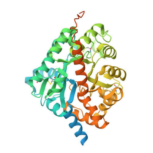
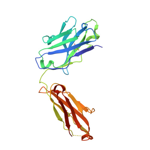
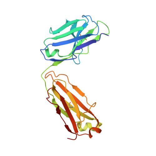
Structure of human DPEP3 in complex with the SC-003 antibody Fab fragment reveals basis for lack of dipeptidase activity.

Hayashi, K.Longenecker, K.L.Koenig, P.Prashar, A.Hampl, J.Stoll, V.Vivona, S.

(2020) J Struct Biol 211: 107512-107512

  • DOI: https://doi.org/10.1016/j.jsb.2020.107512
  • Primary Citation of Related Structures:  
    6VGO, 6VGR

  • PubMed Abstract: 

    Dipeptidase 3 (DPEP3) is one of three glycosylphosphatidylinositol-anchored metallopeptidases potentially involved in the hydrolytic metabolism of dipeptides. While its exact biological function is not clear, DPEP3 expression is normally limited to testis, but can be elevated in ovarian cancer. Antibody drug conjugates targeting DPEP3 have shown efficacy in preclinical models with a pyrrolobenzodiazepine conjugate, SC-003, dosed in a phase I clinical trial (NCT02539719). Here we reveal the novel atomic structure of DPEP3 alone and in complex with the SC-003 Fab fragment at 1.8 and 2.8 Å, respectively. The structure of DPEP3/SC-003 Fab complex reveals an eighteen-residue epitope across the DPEP3 dimerization interface distinct from the enzymatic active site. DPEP1 and DPEP3 extracellular domains share a conserved, dimeric TIM (β/α)8-barrel fold, consistent with 49% sequence identity. However, DPEP3 diverges from DPEP1 and DPEP2 in key positions of its active site: a histidine to tyrosine variation at position 269 reduces affinity for the β zinc and may cause substrate steric hindrance, whereas an aspartate to asparagine change at position 359 abolishes activation of the nucleophilic water/hydroxide, resulting in no in vitro activity against a variety of dipeptides and biological substrates (imipenem, leukotriene D4 and cystinyl-bis-glycine). Hence DPEP3, unlike DPEP1 and DPEP2, may require an activating co-factor in vivo or may remain an inactive, degenerate enzyme. This report sheds light on the structural discriminants between active and inactive membrane dipeptidases and provides a benchmark to characterize current and future DPEP3-targeted therapeutic approaches.


  • Organizational Affiliation

    Research and Development, AbbVie Inc, South San Francisco, CA 94080, United States.


Macromolecules
Find similar proteins by:  (by identity cutoff)  |  3D Structure
Entity ID: 1
MoleculeChains Sequence LengthOrganismDetailsImage
Dipeptidase 3
A, B
488Homo sapiensMutation(s): 0 
Gene Names: DPEP3UNQ834/PRO1772
EC: 3.4.13.19
UniProt & NIH Common Fund Data Resources
Find proteins for Q9H4B8 (Homo sapiens)
Explore Q9H4B8 
Go to UniProtKB:  Q9H4B8
PHAROS:  Q9H4B8
GTEx:  ENSG00000141096 
Entity Groups  
Sequence Clusters30% Identity50% Identity70% Identity90% Identity95% Identity100% Identity
UniProt GroupQ9H4B8
Sequence Annotations
Expand
  • Reference Sequence
Find similar proteins by:  (by identity cutoff)  |  3D Structure
Entity ID: 2
MoleculeChains Sequence LengthOrganismDetailsImage
SC-003 Fab Heavy ChainC,
E [auth H]
226Homo sapiensMutation(s): 0 
Entity Groups  
Sequence Clusters30% Identity50% Identity70% Identity90% Identity95% Identity100% Identity
Sequence Annotations
Expand
  • Reference Sequence
Find similar proteins by:  (by identity cutoff)  |  3D Structure
Entity ID: 3
MoleculeChains Sequence LengthOrganismDetailsImage
SC-003 Fab Light ChainD,
F [auth L]
215Homo sapiensMutation(s): 0 
Entity Groups  
Sequence Clusters30% Identity50% Identity70% Identity90% Identity95% Identity100% Identity
Sequence Annotations
Expand
  • Reference Sequence
Experimental Data & Validation

Experimental Data

  • Method: X-RAY DIFFRACTION
  • Resolution: 2.84 Å
  • R-Value Free: 0.248 
  • R-Value Work: 0.190 
  • R-Value Observed: 0.193 
  • Space Group: P 63
Unit Cell:
Length ( Å )Angle ( ˚ )
a = 167.03α = 90
b = 167.03β = 90
c = 116.38γ = 120
Software Package:
Software NamePurpose
BUSTERrefinement
PDB_EXTRACTdata extraction
XDSdata reduction
Aimlessdata scaling
PHASERphasing

Structure Validation

View Full Validation Report

Currently 6VGR does not have a validation slider image.


Entry History 

Deposition Data

Revision History  (Full details and data files)

  • Version 1.0: 2020-05-13
    Type: Initial release
  • Version 1.1: 2020-06-24
    Changes: Database references
  • Version 1.2: 2023-10-11
    Changes: Data collection, Database references, Refinement description