6V82

Crystal structure of tryptophan synthase from Chlamydia trachomatis D/UW-3/CX


Experimental Data Snapshot

  • Method: X-RAY DIFFRACTION
  • Resolution: 2.42 Å
  • R-Value Free: 0.194 
  • R-Value Work: 0.159 
  • R-Value Observed: 0.161 

wwPDB Validation   3D Report Full Report


This is version 1.4 of the entry. See complete history


Literature

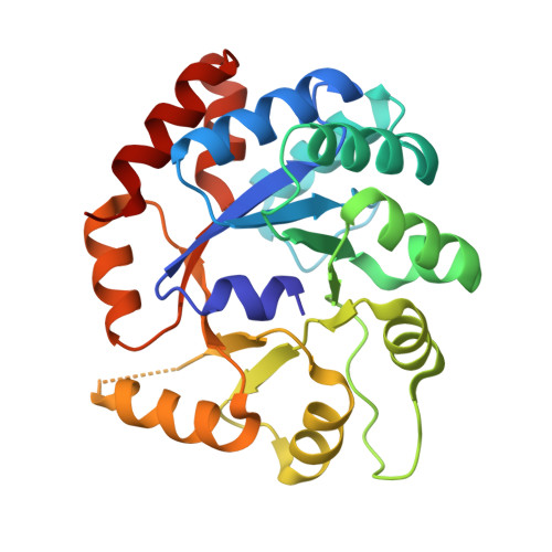
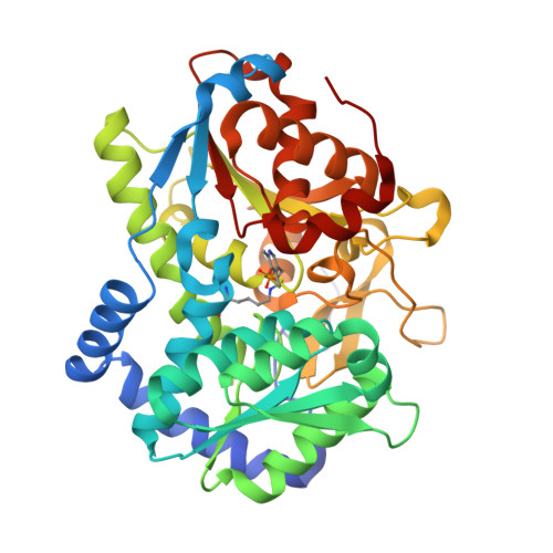
Catalytically impaired TrpA subunit of tryptophan synthase from Chlamydia trachomatis is an allosteric regulator of TrpB.

Michalska, K.Wellington, S.Maltseva, N.Jedrzejczak, R.Selem-Mojica, N.Rosas-Becerra, L.R.Barona-Gomez, F.Hung, D.T.Joachimiak, A.

(2021) Protein Sci 30: 1904-1918

  • DOI: https://doi.org/10.1002/pro.4143
  • Primary Citation of Related Structures:  
    6V82

  • PubMed Abstract: 

    Intracellular growth and pathogenesis of Chlamydia species is controlled by the availability of tryptophan, yet the complete biosynthetic pathway for l-Trp is absent among members of the genus. Some representatives, however, preserve genes encoding tryptophan synthase, TrpAB - a bifunctional enzyme catalyzing the last two steps in l-Trp synthesis. TrpA (subunit α) converts indole-3-glycerol phosphate into indole and glyceraldehyde-3-phosphate (α reaction). The former compound is subsequently used by TrpB (subunit β) to produce l-Trp in the presence of l-Ser and a pyridoxal 5'-phosphate cofactor (β reaction). Previous studies have indicated that in Chlamydia, TrpA has lost its catalytic activity yet remains associated with TrpB to support the β reaction. Here, we provide detailed analysis of the TrpAB from C. trachomatis D/UW-3/CX, confirming that accumulation of mutations in the active site of TrpA renders it enzymatically inactive, despite the conservation of the catalytic residues. We also show that TrpA remains a functional component of the TrpAB complex, increasing the activity of TrpB by four-fold. The side chain of non-conserved βArg267 functions as cation effector, potentially rendering the enzyme less susceptible to the solvent ion composition. The observed structural and functional changes detected herein were placed in a broader evolutionary and genomic context, allowing identification of these mutations in relation to their trp gene contexts in which they occur. Moreover, in agreement with the in vitro data, partial relaxation of purifying selection for TrpA, but not for TrpB, was detected, reinforcing a partial loss of TrpA functions during the course of evolution.


  • Organizational Affiliation

    Center for Structural Genomics of Infectious Diseases, University of Chicago, Chicago, Illinois, USA.


Macromolecules
Find similar proteins by:  (by identity cutoff)  |  3D Structure
Entity ID: 1
MoleculeChains Sequence LengthOrganismDetailsImage
Tryptophan synthase alpha chain253Chlamydia trachomatis D/UW-3/CXMutation(s): 0 
Gene Names: trpACT_171
EC: 4.2.1.20
UniProt
Find proteins for O84173 (Chlamydia trachomatis (strain D/UW-3/Cx))
Explore O84173 
Go to UniProtKB:  O84173
Entity Groups  
Sequence Clusters30% Identity50% Identity70% Identity90% Identity95% Identity100% Identity
UniProt GroupO84173
Sequence Annotations
Expand
  • Reference Sequence
Find similar proteins by:  (by identity cutoff)  |  3D Structure
Entity ID: 2
MoleculeChains Sequence LengthOrganismDetailsImage
Tryptophan synthase beta chain392Chlamydia trachomatis D/UW-3/CXMutation(s): 0 
Gene Names: trpBCT_170
EC: 4.2.1.20
UniProt
Find proteins for O84172 (Chlamydia trachomatis (strain D/UW-3/Cx))
Explore O84172 
Go to UniProtKB:  O84172
Entity Groups  
Sequence Clusters30% Identity50% Identity70% Identity90% Identity95% Identity100% Identity
UniProt GroupO84172
Sequence Annotations
Expand
  • Reference Sequence
Oligosaccharides

Help

Entity ID: 3
MoleculeChains Length2D Diagram Glycosylation3D Interactions
beta-D-fructofuranose-(2-1)-alpha-D-glucopyranose
C, D
2N/A
Glycosylation Resources
GlyTouCan:  G05551OP
GlyCosmos:  G05551OP
Small Molecules
Modified Residues  1 Unique
IDChains TypeFormula2D DiagramParent
LLP
Query on LLP
B
L-PEPTIDE LINKINGC14 H22 N3 O7 PLYS
Biologically Interesting Molecules (External Reference) 1 Unique
Experimental Data & Validation

Experimental Data

  • Method: X-RAY DIFFRACTION
  • Resolution: 2.42 Å
  • R-Value Free: 0.194 
  • R-Value Work: 0.159 
  • R-Value Observed: 0.161 
  • Space Group: C 2 2 21
Unit Cell:
Length ( Å )Angle ( ˚ )
a = 114.923α = 90
b = 133.901β = 90
c = 128.428γ = 90
Software Package:
Software NamePurpose
PHENIXrefinement
HKL-3000data reduction
HKL-3000data scaling
PHENIXphasing

Structure Validation

View Full Validation Report


Entry History & Funding Information

Deposition Data


Funding OrganizationLocationGrant Number
National Institutes of Health/National Institute Of Allergy and Infectious Diseases (NIH/NIAID)United StatesHHSN272201700060C

Revision History  (Full details and data files)

  • Version 1.0: 2020-12-16
    Type: Initial release
  • Version 1.1: 2021-06-30
    Changes: Database references
  • Version 1.2: 2021-09-01
    Changes: Database references
  • Version 1.3: 2023-10-11
    Changes: Data collection, Refinement description
  • Version 1.4: 2023-11-15
    Changes: Data collection