6V3J

KIR3DL1 in complex with HLA-B*57:03 presenting the peptide LSSPVTKSF


Experimental Data Snapshot

  • Method: X-RAY DIFFRACTION
  • Resolution: 1.98 Å
  • R-Value Free: 0.236 
  • R-Value Work: 0.191 
  • R-Value Observed: 0.193 

wwPDB Validation   3D Report Full Report

Currently 6V3J does not have a validation slider image.


This is version 1.4 of the entry. See complete history


Literature

The molecular basis of how buried human leukocyte antigen polymorphism modulates natural killer cell function.

Saunders, P.M.MacLachlan, B.J.Pymm, P.Illing, P.T.Deng, Y.Wong, S.C.Oates, C.V.L.Purcell, A.W.Rossjohn, J.Vivian, J.P.Brooks, A.G.

(2020) Proc Natl Acad Sci U S A 117: 11636-11647

  • DOI: https://doi.org/10.1073/pnas.1920570117
  • Primary Citation of Related Structures:  
    6V2O, 6V2P, 6V2Q, 6V3J

  • PubMed Abstract: 

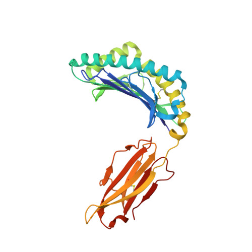
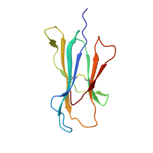
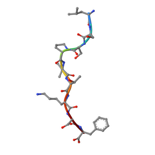
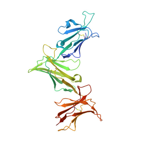
    Micropolymorphisms within human leukocyte antigen (HLA) class I molecules can change the architecture of the peptide-binding cleft, leading to differences in peptide presentation and T cell recognition. The impact of such HLA variation on natural killer (NK) cell recognition remains unclear. Given the differential association of HLA-B*57:01 and HLA-B*57:03 with the control of HIV, recognition of these HLA-B57 allomorphs by the killer cell immunoglobulin-like receptor (KIR) 3DL1 was compared. Despite differing by only two polymorphic residues, both buried within the peptide-binding cleft, HLA-B*57:01 more potently inhibited NK cell activation. Direct-binding studies showed KIR3DL1 to preferentially recognize HLA-B*57:01, particularly when presenting peptides with positively charged position (P)Ω-2 residues. In HLA-B*57:01, charged PΩ-2 residues were oriented toward the peptide-binding cleft and away from KIR3DL1. In HLA-B*57:03, the charged PΩ-2 residues protruded out from the cleft and directly impacted KIR3DL1 engagement. Accordingly, KIR3DL1 recognition of HLA class I ligands is modulated by both the peptide sequence and conformation, as determined by the HLA polymorphic framework, providing a rationale for understanding differences in clinical associations.


  • Organizational Affiliation

    Department of Microbiology and Immunology, Peter Doherty Institute for Infection and Immunity, The University of Melbourne, Melbourne, VIC 3010, Australia.


Macromolecules
Find similar proteins by:  (by identity cutoff)  |  3D Structure
Entity ID: 1
MoleculeChains Sequence LengthOrganismDetailsImage
HLA-B alpha chain (B*5703GB)275Homo sapiensMutation(s): 0 
Gene Names: HLA-BHLA-B*5703GB
UniProt
Find proteins for I3ZN84 (Homo sapiens)
Explore I3ZN84 
Go to UniProtKB:  I3ZN84
Entity Groups  
Sequence Clusters30% Identity50% Identity70% Identity90% Identity95% Identity100% Identity
UniProt GroupI3ZN84
Sequence Annotations
Expand
  • Reference Sequence
Find similar proteins by:  (by identity cutoff)  |  3D Structure
Entity ID: 2
MoleculeChains Sequence LengthOrganismDetailsImage
Beta-2-microglobulin100Homo sapiensMutation(s): 0 
Gene Names: B2MCDABP0092HDCMA22P
UniProt & NIH Common Fund Data Resources
Find proteins for P61769 (Homo sapiens)
Explore P61769 
Go to UniProtKB:  P61769
PHAROS:  P61769
GTEx:  ENSG00000166710 
Entity Groups  
Sequence Clusters30% Identity50% Identity70% Identity90% Identity95% Identity100% Identity
UniProt GroupP61769
Sequence Annotations
Expand
  • Reference Sequence

Find similar proteins by:  Sequence   |   3D Structure  

Entity ID: 3
MoleculeChains Sequence LengthOrganismDetailsImage
Peptide Leu-Ser-Ser-Pro-Val-Thr-Lys-Ser-Phe9synthetic constructMutation(s): 0 
Sequence Annotations
Expand
  • Reference Sequence
Find similar proteins by:  (by identity cutoff)  |  3D Structure
Entity ID: 4
MoleculeChains Sequence LengthOrganismDetailsImage
Killer cell immunoglobulin-like receptor 3DL1D [auth G]299Homo sapiensMutation(s): 0 
Gene Names: KIR3DL1CD158ENKAT3NKB1
UniProt & NIH Common Fund Data Resources
Find proteins for P43629 (Homo sapiens)
Explore P43629 
Go to UniProtKB:  P43629
PHAROS:  P43629
GTEx:  ENSG00000167633 
Entity Groups  
Sequence Clusters30% Identity50% Identity70% Identity90% Identity95% Identity100% Identity
UniProt GroupP43629
Sequence Annotations
Expand
  • Reference Sequence
Experimental Data & Validation

Experimental Data

  • Method: X-RAY DIFFRACTION
  • Resolution: 1.98 Å
  • R-Value Free: 0.236 
  • R-Value Work: 0.191 
  • R-Value Observed: 0.193 
  • Space Group: P 1
Unit Cell:
Length ( Å )Angle ( ˚ )
a = 51.759α = 96.65
b = 60.771β = 97.39
c = 65.36γ = 109.18
Software Package:
Software NamePurpose
PHENIXrefinement
XDSdata reduction
Aimlessdata scaling
PHASERphasing

Structure Validation

View Full Validation Report

Currently 6V3J does not have a validation slider image.


Entry History 

Deposition Data

Revision History  (Full details and data files)

  • Version 1.0: 2020-05-20
    Type: Initial release
  • Version 1.1: 2020-05-27
    Changes: Database references
  • Version 1.2: 2020-06-10
    Changes: Database references
  • Version 1.3: 2020-07-29
    Type: Remediation
    Reason: Carbohydrate remediation
    Changes: Data collection, Derived calculations, Structure summary
  • Version 1.4: 2023-10-11
    Changes: Data collection, Database references, Refinement description, Structure summary