


Crystal structure of malate dehydrogenase from Naegleria fowleri ATCC 30863
Abendroth, J., Lorimer, D.D., Horanyi, P.S., Edwards, T.E.To be published.
Experimental Data Snapshot
wwPDB Validation 3D Report Full Report

Entity ID: 1 | |||||
|---|---|---|---|---|---|
| Molecule | Chains | Sequence Length | Organism | Details | Image |
| Malate dehydrogenase | 444 | Naegleria fowleri | Mutation(s): 0 Gene Names: mRNA1_NF0021050 | 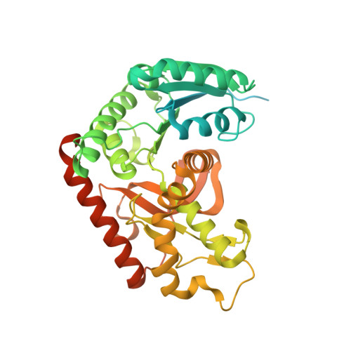 | |
Entity Groups | |||||
| Sequence Clusters | 30% Identity50% Identity70% Identity90% Identity95% Identity100% Identity | ||||
Sequence AnnotationsExpand | |||||
| |||||
| Ligands 1 Unique | |||||
|---|---|---|---|---|---|
| ID | Chains | Name / Formula / InChI Key | 2D Diagram | 3D Interactions | |
| EDO Query on EDO | M [auth D], N [auth J], O [auth J], P [auth L] | 1,2-ETHANEDIOL C2 H6 O2 LYCAIKOWRPUZTN-UHFFFAOYSA-N |  | ||
| Length ( Å ) | Angle ( ˚ ) |
|---|---|
| a = 60.33 | α = 91.093 |
| b = 137.48 | β = 89.998 |
| c = 139.28 | γ = 91.482 |
| Software Name | Purpose |
|---|---|
| PHENIX | refinement |
| XDS | data reduction |
| XSCALE | data scaling |
| PDB_EXTRACT | data extraction |
| MoRDa | phasing |
| ARP/wARP | model building |
| BUCCANEER | model building |
RCSB PDB (citation) is hosted by
RCSB PDB is a member of the

