6UH1

Structure of the EVA71 strain 11316 capsid


Experimental Data Snapshot

  • Method: ELECTRON MICROSCOPY
  • Resolution: 3.04 Å
  • Aggregation State: PARTICLE 
  • Reconstruction Method: SINGLE PARTICLE 

wwPDB Validation   3D Report Full Report


This is version 1.2 of the entry. See complete history


Literature

Scaffold Simplification Strategy Leads to a Novel Generation of Dual Human Immunodeficiency Virus and Enterovirus-A71 Entry Inhibitors.

Martinez-Gualda, B.Sun, L.Marti-Mari, O.Noppen, S.Abdelnabi, R.Bator, C.M.Quesada, E.Delang, L.Mirabelli, C.Lee, H.Schols, D.Neyts, J.Hafenstein, S.Camarasa, M.J.Gago, F.San-Felix, A.

(2020) J Med Chem 63: 349-368

  • DOI: https://doi.org/10.1021/acs.jmedchem.9b01737
  • Primary Citation of Related Structures:  
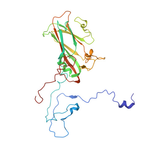
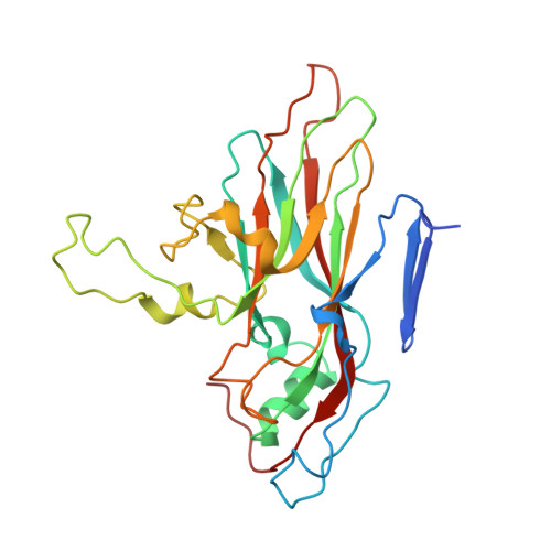
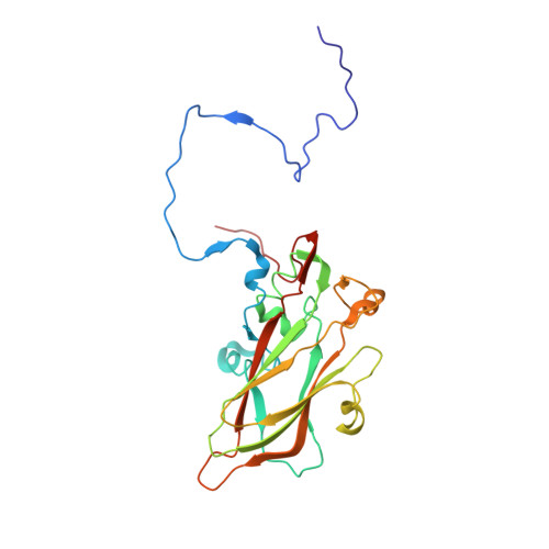
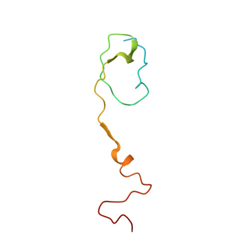
    6UH1, 6UH6, 6UH7

  • PubMed Abstract: 

    Currently, there are only three FDA-approved drugs that inhibit human immunodeficiency virus (HIV) entry-fusion into host cells. The situation is even worse for enterovirus EV71 infection for which no antiviral therapies are available. We describe here the discovery of potent entry dual inhibitors of HIV and EV71. These compounds contain in their structure three or four tryptophan (Trp) residues linked to a central scaffold. Critical for anti-HIV/EV71 activity is the presence of extra phenyl rings, bearing one or two carboxylates, at the C2 position of the indole ring of each Trp residue. The most potent derivatives, 22 and 30 , inhibit early steps of the replicative cycles of HIV-1 and EV-A71 by interacting with their respective viral surfaces (glycoprotein gp120 of HIV and the fivefold axis of the EV-A71 capsid). The high potency, low toxicity, facile chemical synthesis, and great opportunities for chemical optimization make them useful prototypes for future medicinal chemistry studies.


  • Organizational Affiliation

    Instituto de Química Médica (IQM-CSIC) , 28006 Madrid , Spain.


Macromolecules
Find similar proteins by:  (by identity cutoff)  |  3D Structure
Entity ID: 1
MoleculeChains Sequence LengthOrganismDetailsImage
VP1297Enterovirus A71Mutation(s): 0 
UniProt
Find proteins for D4QGA8 (Human enterovirus 71)
Explore D4QGA8 
Go to UniProtKB:  D4QGA8
Entity Groups  
Sequence Clusters30% Identity50% Identity70% Identity90% Identity95% Identity100% Identity
UniProt GroupD4QGA8
Sequence Annotations
Expand
  • Reference Sequence
Find similar proteins by:  (by identity cutoff)  |  3D Structure
Entity ID: 2
MoleculeChains Sequence LengthOrganismDetailsImage
VP2254Enterovirus A71Mutation(s): 0 
UniProt
Find proteins for I6W7A3 (Human enterovirus 71)
Explore I6W7A3 
Go to UniProtKB:  I6W7A3
Entity Groups  
Sequence Clusters30% Identity50% Identity70% Identity90% Identity95% Identity100% Identity
UniProt GroupI6W7A3
Sequence Annotations
Expand
  • Reference Sequence
Find similar proteins by:  (by identity cutoff)  |  3D Structure
Entity ID: 3
MoleculeChains Sequence LengthOrganismDetailsImage
VP3242Enterovirus A71Mutation(s): 0 
EC: 3.4.22.29 (PDB Primary Data), 3.6.1.15 (PDB Primary Data), 3.4.22.28 (PDB Primary Data), 2.7.7.48 (PDB Primary Data)
UniProt
Find proteins for A0A0E3SXU7 (Human enterovirus 71)
Explore A0A0E3SXU7 
Go to UniProtKB:  A0A0E3SXU7
Entity Groups  
Sequence Clusters30% Identity50% Identity70% Identity90% Identity95% Identity100% Identity
UniProt GroupA0A0E3SXU7
Sequence Annotations
Expand
  • Reference Sequence
Find similar proteins by:  (by identity cutoff)  |  3D Structure
Entity ID: 4
MoleculeChains Sequence LengthOrganismDetailsImage
VP469Enterovirus A71Mutation(s): 0 
EC: 3.4.22.29 (PDB Primary Data), 3.6.1.15 (PDB Primary Data), 3.4.22.28 (PDB Primary Data), 2.7.7.48 (PDB Primary Data)
UniProt
Find proteins for E9RGA0 (Human enterovirus 71)
Explore E9RGA0 
Go to UniProtKB:  E9RGA0
Entity Groups  
Sequence Clusters30% Identity50% Identity70% Identity90% Identity95% Identity100% Identity
UniProt GroupE9RGA0
Sequence Annotations
Expand
  • Reference Sequence
Small Molecules
Ligands 1 Unique
IDChains Name / Formula / InChI Key2D Diagram3D Interactions
SPH
Query on SPH

Download Ideal Coordinates CCD File 
E [auth A]SPHINGOSINE
C18 H37 N O2
WWUZIQQURGPMPG-MSOLQXFVSA-N
Experimental Data & Validation

Experimental Data

  • Method: ELECTRON MICROSCOPY
  • Resolution: 3.04 Å
  • Aggregation State: PARTICLE 
  • Reconstruction Method: SINGLE PARTICLE 

Structure Validation

View Full Validation Report


Entry History 

Deposition Data

Revision History  (Full details and data files)

  • Version 1.0: 2019-12-18
    Type: Initial release
  • Version 1.1: 2020-01-22
    Changes: Database references
  • Version 1.2: 2024-03-20
    Changes: Data collection, Database references, Derived calculations