6U2F

PCSK9-Fab 7G7 complex bound to cis-1-amino-4-phenylcyclohexaneacyl-WNLK(hR)IGLLR - NH2


Experimental Data Snapshot

  • Method: X-RAY DIFFRACTION
  • Resolution: 2.94 Å
  • R-Value Free: 0.209 
  • R-Value Work: 0.174 
  • R-Value Observed: 0.175 

wwPDB Validation   3D Report Full Report


This is version 2.0 of the entry. See complete history


Literature

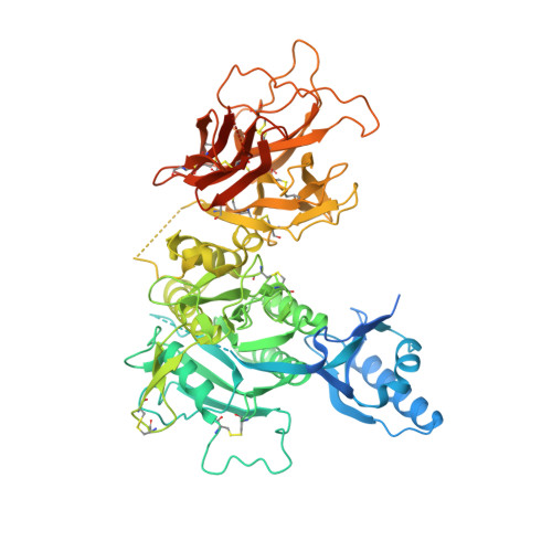
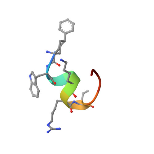
Design of Organo-Peptides As Bipartite PCSK9 Antagonists.

Burdick, D.J.Skelton, N.J.Ultsch, M.Beresini, M.H.Eigenbrot, C.Li, W.Zhang, Y.Nguyen, H.Kong-Beltran, M.Quinn, J.G.Kirchhofer, D.

(2020) ACS Chem Biol 15: 425-436

  • DOI: https://doi.org/10.1021/acschembio.9b00899
  • Primary Citation of Related Structures:  
    6U2F, 6U3I

  • PubMed Abstract: 

    Proprotein convertase subtilisin/kexin 9 (PCSK9) has become an important therapeutic target for lipid lowering, since it regulates low-density lipoprotein cholesterol (LDL-c) levels by binding to liver LDL receptors (LDLR) and effecting their intracellular degradation. However, the development of small molecule inhibitors is hampered by the lack of attractive PCSK9 target sites. We recently discovered helical peptides that are able to bind to a cryptic groove site on PCSK9, which is situated in proximity to the main LDLR binding site. Here, we designed potent bipartite PCSK9 inhibitors by appending organic moieties to a helical groove-binding peptide to reach a hydrophobic pocket in the proximal LDLR binding region. The ultimately designed 1-amino-4-phenylcyclohexane-1-carbonyl extension improved the peptide affinity by >100-fold, yielding organo-peptide antagonists that potently inhibited PCSK9 binding to LDLR and preserved cellular LDLR. These new bipartite antagonists have reduced mass and improved potency compared to the first-generation peptide antagonists, further validating the PCSK9 groove as a viable therapeutic target site.


Macromolecules
Find similar proteins by:  (by identity cutoff)  |  3D Structure
Entity ID: 1
MoleculeChains Sequence LengthOrganismDetailsImage
Proprotein convertase subtilisin/kexin type 9700Homo sapiensMutation(s): 2 
Gene Names: PCSK9NARC1PSEC0052
EC: 3.4.21
UniProt & NIH Common Fund Data Resources
Find proteins for Q8NBP7 (Homo sapiens)
Explore Q8NBP7 
Go to UniProtKB:  Q8NBP7
PHAROS:  Q8NBP7
GTEx:  ENSG00000169174 
Entity Groups  
Sequence Clusters30% Identity50% Identity70% Identity90% Identity95% Identity100% Identity
UniProt GroupQ8NBP7
Sequence Annotations
Expand
  • Reference Sequence

Find similar proteins by:  Sequence   |   3D Structure  

Entity ID: 2
MoleculeChains Sequence LengthOrganismDetailsImage
Organo-peptide PCSK9 inhibitor11synthetic constructMutation(s): 0 
Entity Groups  
Sequence Clusters30% Identity50% Identity70% Identity90% Identity95% Identity100% Identity
Sequence Annotations
Expand
  • Reference Sequence
Find similar proteins by:  (by identity cutoff)  |  3D Structure
Entity ID: 3
MoleculeChains Sequence LengthOrganismDetailsImage
7G7 heavy chainC [auth H]223Mus musculusMutation(s): 0 
UniProt
Find proteins for Q569X1 (Mus musculus)
Explore Q569X1 
Go to UniProtKB:  Q569X1
Entity Groups  
Sequence Clusters30% Identity50% Identity70% Identity90% Identity95% Identity100% Identity
UniProt GroupQ569X1
Sequence Annotations
Expand
  • Reference Sequence
Find similar proteins by:  (by identity cutoff)  |  3D Structure
Entity ID: 4
MoleculeChains Sequence LengthOrganismDetailsImage
7G7 light chainD [auth L]214Mus musculusMutation(s): 0 
UniProt
Find proteins for A0A0U5BC76 (Mus musculus)
Explore A0A0U5BC76 
Go to UniProtKB:  A0A0U5BC76
Entity Groups  
Sequence Clusters30% Identity50% Identity70% Identity90% Identity95% Identity100% Identity
UniProt GroupA0A0U5BC76
Sequence Annotations
Expand
  • Reference Sequence
Small Molecules
Ligands 1 Unique
IDChains Name / Formula / InChI Key2D Diagram3D Interactions
CA
Query on CA

Download Ideal Coordinates CCD File 
E [auth A]CALCIUM ION
Ca
BHPQYMZQTOCNFJ-UHFFFAOYSA-N
Modified Residues  1 Unique
IDChains TypeFormula2D DiagramParent
HRG
Query on HRG
B
L-PEPTIDE LINKINGC7 H16 N4 O2ARG
Biologically Interesting Molecules (External Reference) 1 Unique
Experimental Data & Validation

Experimental Data

  • Method: X-RAY DIFFRACTION
  • Resolution: 2.94 Å
  • R-Value Free: 0.209 
  • R-Value Work: 0.174 
  • R-Value Observed: 0.175 
  • Space Group: I 2 2 2
Unit Cell:
Length ( Å )Angle ( ˚ )
a = 110.569α = 90
b = 142.74β = 90
c = 239.287γ = 90
Software Package:
Software NamePurpose
BUSTERrefinement
PDB_EXTRACTdata extraction
HKL-2000data reduction
SCALEPACKdata scaling
PHASERphasing

Structure Validation

View Full Validation Report


Entry History 

Deposition Data

Revision History  (Full details and data files)

  • Version 1.0: 2020-02-05
    Type: Initial release
  • Version 1.1: 2020-03-04
    Changes: Database references
  • Version 1.2: 2020-08-12
    Changes: Derived calculations, Structure summary
  • Version 1.3: 2023-10-11
    Changes: Data collection, Database references, Refinement description
  • Version 2.0: 2023-11-15
    Changes: Atomic model, Data collection, Derived calculations