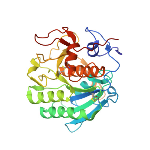
6TXG

Proteinase K in complex with a "half sandwich"-type Ru(II) coordination compound


Experimental Data Snapshot

  • Method: X-RAY DIFFRACTION
  • Resolution: 1.37 Å
  • R-Value Free: 0.168 
  • R-Value Work: 0.151 
  • R-Value Observed: 0.152 

wwPDB Validation   3D Report Full Report


Ligand Structure Quality Assessment 


This is version 1.2 of the entry. See complete history


Literature

High-resolution crystal structures of a "half sandwich"-type Ru(II) coordination compound bound to hen egg-white lysozyme and proteinase K.

Chiniadis, L.Bratsos, I.Bethanis, K.Karpusas, M.Giastas, P.Papakyriakou, A.

(2020) J Biol Inorg Chem 25: 635-645

  • DOI: https://doi.org/10.1007/s00775-020-01786-z
  • Primary Citation of Related Structures:  
    6TVL, 6TXG

  • PubMed Abstract: 

    The high-resolution X-ray crystal structures of the adducts formed between the "half sandwich"-type Ru(II) coordination compound [Ru II (1,4,7-trithiacyclononane)(ethane-1,2-diamine)Cl] + and two proteins, namely hen egg-white lysozyme and proteinase K, are presented. The structures unveil that upon reaction with both enzymes the Ru(II) compound is coordinated by solvent-exposed aspartate residues after releasing the chloride ligand (Asp101 in lysozyme, Asp200 and Asp260 in proteinase K), while retaining the two chelating ligands. The adduct with Asp101 residue at the catalytic cleft of lysozyme is accompanied by residue-specific conformational changes to accommodate the Ru(II) fragment, whereas the complexes bound at the two calcium-binding sites of proteinase K revealed minimal structural perturbation of the enzyme. To the best of our knowledge, proteinase K is used here for the first time as a model system of protein metalation and these are the first X-ray crystal structures of protein adducts of a Ru(II) coordination compound that maintains its coordination sphere almost intact upon binding. Our data demonstrate the role of ligands in stabilizing the protein adducts via hydrophobic/aromatic or hydrogen-bonding interactions, as well as their underlying role in the selection of specific sites on the electrostatic potential surface of the enzymes.


  • Organizational Affiliation

    Institute of Biosciences and Applications, National Centre for Scientific Research "Demokritos", 15341, Athens, Greece.


Macromolecules
Find similar proteins by:  (by identity cutoff)  |  3D Structure
Entity ID: 1
MoleculeChains Sequence LengthOrganismDetailsImage
Proteinase K279Parengyodontium albumMutation(s): 0 
EC: 3.4.21.64
UniProt
Find proteins for P06873 (Parengyodontium album)
Explore P06873 
Go to UniProtKB:  P06873
Entity Groups  
Sequence Clusters30% Identity50% Identity70% Identity90% Identity95% Identity100% Identity
UniProt GroupP06873
Sequence Annotations
Expand
  • Reference Sequence
Small Molecules
Ligands 6 Unique
IDChains Name / Formula / InChI Key2D Diagram3D Interactions
NYN (Subject of Investigation/LOI)
Query on NYN

Download Ideal Coordinates CCD File 
H [auth A],
J [auth A]
chlorido(1,2-diaminoethane-k2N,N')(1,4,7-trithiacyclononane-k3S,S',S'')ruthenium(II) trifluoromethanesulfonate
C8 H16 N2 Ru S3
ZNOIBYRQMPADCX-UHFFFAOYSA-N
TFS
Query on TFS

Download Ideal Coordinates CCD File 
K [auth A]trifluoromethanesulfonic acid
C H F3 O3 S
ITMCEJHCFYSIIV-UHFFFAOYSA-N
EDO
Query on EDO

Download Ideal Coordinates CCD File 
E [auth A],
F [auth A]
1,2-ETHANEDIOL
C2 H6 O2
LYCAIKOWRPUZTN-UHFFFAOYSA-N
NO3
Query on NO3

Download Ideal Coordinates CCD File 
B [auth A],
C [auth A],
D [auth A]
NITRATE ION
N O3
NHNBFGGVMKEFGY-UHFFFAOYSA-N
CA
Query on CA

Download Ideal Coordinates CCD File 
G [auth A]CALCIUM ION
Ca
BHPQYMZQTOCNFJ-UHFFFAOYSA-N
NA
Query on NA

Download Ideal Coordinates CCD File 
I [auth A]SODIUM ION
Na
FKNQFGJONOIPTF-UHFFFAOYSA-N
Experimental Data & Validation

Experimental Data

  • Method: X-RAY DIFFRACTION
  • Resolution: 1.37 Å
  • R-Value Free: 0.168 
  • R-Value Work: 0.151 
  • R-Value Observed: 0.152 
  • Space Group: P 43 21 2
Unit Cell:
Length ( Å )Angle ( ˚ )
a = 67.708α = 90
b = 67.708β = 90
c = 107.314γ = 90
Software Package:
Software NamePurpose
PHENIXrefinement
PDB_EXTRACTdata extraction
XDSdata reduction
XDSdata scaling
PHASERphasing

Structure Validation

View Full Validation Report


Ligand Structure Quality Assessment 


Entry History 

Deposition Data

Revision History  (Full details and data files)

  • Version 1.0: 2020-04-29
    Type: Initial release
  • Version 1.1: 2020-06-03
    Changes: Database references
  • Version 1.2: 2024-01-24
    Changes: Data collection, Database references, Derived calculations, Refinement description