Reverse protein engineering of a novel 4-domain copper nitrite reductase reveals functional regulation by protein-protein interaction.
Sasaki, D., Watanabe, T.F., Eady, R.R., Garratt, R.C., Antonyuk, S.V., Hasnain, S.S.(2021) FEBS J 288: 262-280
- PubMed: 32255260
- DOI: https://doi.org/10.1111/febs.15324
- Primary Citation of Related Structures:
6THE, 6THF - PubMed Abstract:
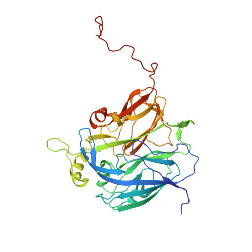
Cu-containing nitrite reductases that convert NO 2 - to NO are critical enzymes in nitrogen-based energy metabolism. Among organisms in the order Rhizobiales, we have identified two copies of nirK, one encoding a new class of 4-domain CuNiR that has both cytochrome and cupredoxin domains fused at the N terminus and the other, a classical 2-domain CuNiR (Br 2D NiR). We report the first enzymatic studies of a novel 4-domain CuNiR from Bradyrhizobium sp. ORS 375 (BrNiR), its genetically engineered 3- and 2-domain variants, and Br 2D NiR revealing up to ~ 500-fold difference in catalytic efficiency in comparison with classical 2-domain CuNiRs. Contrary to the expectation that tethering would enhance electron delivery by restricting the conformational search by having a self-contained donor-acceptor system, we demonstrate that 4-domain BrNiR utilizes N-terminal tethering for downregulating enzymatic activity instead. Both Br 2D NiR and an engineered 2-domain variant of BrNiR (Δ(Cytc-Cup) BrNiR) have 3 to 5% NiR activity compared to the well-characterized 2-domain CuNiRs from Alcaligenes xylosoxidans (AxNiR) and Achromobacter cycloclastes (AcNiR). Structural comparison of Δ(Cytc-Cup) BrNiR and Br 2D NiR with classical 2-domain AxNiR and AcNiR reveals structural differences of the proton transfer pathway that could be responsible for the lowering of activity. Our study provides insights into unique structural and functional characteristics of naturally occurring 4-domain CuNiR and its engineered 3- and 2-domain variants. The reverse protein engineering approach utilized here has shed light onto the broader question of the evolution of transient encounter complexes and tethered electron transfer complexes. ENZYME: Copper-containing nitrite reductase (CuNiR) (EC 1.7.2.1). DATABASE: The atomic coordinate and structure factor of Δ(Cytc-Cup) BrNiR and Br 2D NiR have been deposited in the Protein Data Bank (http://www.rcsb.org/) under the accession code 6THE and 6THF, respectively.
Organizational Affiliation:
Molecular Biophysics Group, Institute of Systems, Molecular and Integrative Biology, Faculty of Health and Life Sciences, University of Liverpool, UK.


