6S2I

Anti-tumor antibody 14F7-derived scFv in complex with NeuGc Gm3


Experimental Data Snapshot

  • Method: X-RAY DIFFRACTION
  • Resolution: 2.29 Å
  • R-Value Free: 0.255 
  • R-Value Work: 0.222 
  • R-Value Observed: 0.224 

wwPDB Validation   3D Report Full Report


This is version 2.2 of the entry. See complete history


Literature

Key role of a structural water molecule for the specificity of 14F7-An antitumor antibody targeting the NeuGc GM3 ganglioside.

Bjerregaard-Andersen, K.Abraha, F.Johannesen, H.Oscarson, S.Moreno, E.Krengel, U.

(2021) Glycobiology 31: 1500-1509

  • DOI: https://doi.org/10.1093/glycob/cwab076
  • Primary Citation of Related Structures:  
    6S2I

  • PubMed Abstract: 

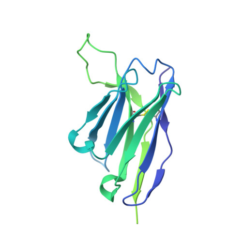
    Tumor-associated glycolipids such as NeuGc GM3 are auspicious molecular targets in antineoplastic therapies and vaccine strategies. 14F7 is a monoclonal IgG1 with high clinical potential in cancer immunotherapy as it displays extraordinary specificity for NeuGc GM3, while it does not recognize the very similar, ubiquitous NeuAc GM3. Here we present the 2.3 Å crystal structure of the 14F7 antigen-binding domain (14F7 scFv) in complex with the NeuGc GM3 trisaccharide. Modeling analysis and previous mutagenesis data suggest that 14F7 may also bind to an alternative NeuGc GM3 conformation, not observed in the crystal structure. The most intriguing finding, however, was that a water molecule centrally placed in the complementarity-determining region directly mediates the specificity of 14F7 to NeuGc GM3. This has profound impact on the complexity of engineering in the binding site and provides an excellent example of the importance in understanding the water structure in antibody-antigen interactions.


  • Organizational Affiliation

    Department of Chemistry, University of Oslo, NO-0315 Oslo, Norway.


Macromolecules
Find similar proteins by:  (by identity cutoff)  |  3D Structure
Entity ID: 1
MoleculeChains Sequence LengthOrganismDetailsImage
scFv C1
A, B, C, D, E
A, B, C, D, E, F, G, H
253Mus musculusMutation(s): 0 
Entity Groups  
Sequence Clusters30% Identity50% Identity70% Identity90% Identity95% Identity100% Identity
Sequence Annotations
Expand
  • Reference Sequence
Oligosaccharides

Help

Entity ID: 2
MoleculeChains Length2D Diagram Glycosylation3D Interactions
N-glycolyl-alpha-neuraminic acid-(2-3)-beta-D-galactopyranose-(1-4)-beta-D-glucopyranoseI [auth J]3N/A
Glycosylation Resources
GlyTouCan:  G75789AR
GlyCosmos:  G75789AR
GlyGen:  G75789AR
Experimental Data & Validation

Experimental Data

  • Method: X-RAY DIFFRACTION
  • Resolution: 2.29 Å
  • R-Value Free: 0.255 
  • R-Value Work: 0.222 
  • R-Value Observed: 0.224 
  • Space Group: P 1 21 1
Unit Cell:
Length ( Å )Angle ( ˚ )
a = 62.88α = 90
b = 113.69β = 91.24
c = 66.95γ = 90
Software Package:
Software NamePurpose
PHENIXrefinement
XDSdata reduction
XSCALEdata scaling
PHENIXphasing

Structure Validation

View Full Validation Report


Entry History 

Deposition Data

Revision History  (Full details and data files)

  • Version 1.0: 2020-07-15
    Type: Initial release
  • Version 2.0: 2021-02-10
    Changes: Atomic model, Data collection, Derived calculations, Non-polymer description, Structure summary
  • Version 2.1: 2022-01-26
    Changes: Data collection, Database references
  • Version 2.2: 2024-01-24
    Changes: Data collection, Refinement description