6RXJ

Crystal structure of CobB wt in complex with H4K16-Acetyl peptide


Experimental Data Snapshot

  • Method: X-RAY DIFFRACTION
  • Resolution: 1.60 Å
  • R-Value Free: 0.188 
  • R-Value Work: 0.173 
  • R-Value Observed: 0.174 

wwPDB Validation   3D Report Full Report


This is version 2.1 of the entry. See complete history


Literature

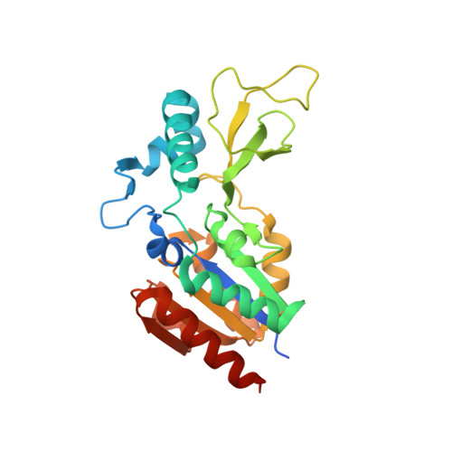
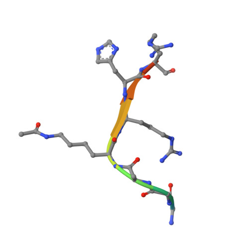
Evolved, Selective Erasers of Distinct Lysine Acylations.

Spinck, M.Neumann-Staubitz, P.Ecke, M.Gasper, R.Neumann, H.

(2020) Angew Chem Int Ed Engl 59: 11142-11149

  • DOI: https://doi.org/10.1002/anie.202002899
  • Primary Citation of Related Structures:  
    6RXJ, 6RXK, 6RXL, 6RXM, 6RXO, 6RXP, 6RXQ, 6RXR, 6RXS

  • PubMed Abstract: 

    Lysine acylations, a family of diverse protein modifications varying in acyl-group length, charge, and saturation, are linked to many important physiological processes. Only a small set of substrate-promiscuous lysine acetyltransferases and deacetylases (KDACs) install and remove this vast variety of modifications. Engineered KDACs that remove only one type of acylation would help to dissect the different contributions of distinct acylations. We developed a bacterial selection system for the directed evolution of KDACs and identified variants up to 400 times more selective for butyryl-lysine compared to crotonyl-lysine. Structural analyses revealed that the enzyme adopts different conformational states depending on the type of acylation of the bound peptide. We used the butyryl-selective KDAC variant to shift the cellular acylation spectrum towards increased lysine crotonylation. These new enzymes will help in dissecting the roles of different lysine acylations in cell physiology.


  • Organizational Affiliation

    Department of Structural Biochemistry, Max-Planck-Institute of Molecular Physiology, Otto-Hahn-Strasse 11, 44227, Dortmund, Germany.


Macromolecules
Find similar proteins by:  (by identity cutoff)  |  3D Structure
Entity ID: 1
MoleculeChains Sequence LengthOrganismDetailsImage
NAD-dependent protein deacylase
A, B
254Escherichia coli K-12Mutation(s): 0 
Gene Names: cobBycfYb1120JW1106
EC: 3.5.1
UniProt
Find proteins for P75960 (Escherichia coli (strain K12))
Explore P75960 
Go to UniProtKB:  P75960
Entity Groups  
Sequence Clusters30% Identity50% Identity70% Identity90% Identity95% Identity100% Identity
UniProt GroupP75960
Sequence Annotations
Expand
  • Reference Sequence

Find similar proteins by:  Sequence   |   3D Structure  

Entity ID: 2
MoleculeChains Sequence LengthOrganismDetailsImage
Histone H4
C, D
10Saccharomyces cerevisiae S288CMutation(s): 0 
UniProt
Find proteins for P02309 (Saccharomyces cerevisiae (strain ATCC 204508 / S288c))
Explore P02309 
Go to UniProtKB:  P02309
Entity Groups  
Sequence Clusters30% Identity50% Identity70% Identity90% Identity95% Identity100% Identity
UniProt GroupP02309
Sequence Annotations
Expand
  • Reference Sequence
Small Molecules
Modified Residues  1 Unique
IDChains TypeFormula2D DiagramParent
ALY
Query on ALY
C, D
L-PEPTIDE LINKINGC8 H16 N2 O3LYS
Experimental Data & Validation

Experimental Data

  • Method: X-RAY DIFFRACTION
  • Resolution: 1.60 Å
  • R-Value Free: 0.188 
  • R-Value Work: 0.173 
  • R-Value Observed: 0.174 
  • Space Group: C 2 2 21
Unit Cell:
Length ( Å )Angle ( ˚ )
a = 131.1α = 90
b = 131.26β = 90
c = 58.54γ = 90
Software Package:
Software NamePurpose
PHENIXrefinement
XDSdata reduction
XSCALEdata scaling
PHASERphasing
PDB_EXTRACTdata extraction

Structure Validation

View Full Validation Report


Entry History & Funding Information

Deposition Data


Funding OrganizationLocationGrant Number
German Research FoundationGermanyNE1589/5-1

Revision History  (Full details and data files)

  • Version 1.0: 2020-04-15
    Type: Initial release
  • Version 1.1: 2020-07-01
    Changes: Database references
  • Version 1.2: 2023-09-27
    Changes: Data collection, Database references, Derived calculations, Refinement description
  • Version 2.0: 2023-11-15
    Changes: Atomic model, Data collection
  • Version 2.1: 2024-01-24
    Changes: Refinement description