6RPR

LEM domain of Emerin mutant T43I in complex with BAF dimer and the Igfold of the lamin A/C

  • Classification: DNA BINDING PROTEIN
  • Organism(s): Homo sapiens
  • Expression System: Escherichia coli
  • Mutation(s): No 

  • Deposited: 2019-05-14 Released: 2020-03-04 
  • Deposition Author(s): Essawy, N., Samson, C.
  • Funding Organization(s): French Infrastructure for Integrated Structural Biology, European Communitys Seventh Framework Programme

Experimental Data Snapshot

  • Method: X-RAY DIFFRACTION
  • Resolution: 2.26 Å
  • R-Value Free: 0.244 
  • R-Value Work: 0.196 
  • R-Value Observed: 0.198 

wwPDB Validation   3D Report Full Report


Ligand Structure Quality Assessment 


This is version 1.1 of the entry. See complete history


Literature

An Emerin LEM-Domain Mutation Impairs Cell Response to Mechanical Stress.

Essawy, N.Samson, C.Petitalot, A.Moog, S.Bigot, A.Herrada, I.Marcelot, A.Arteni, A.A.Coirault, C.Zinn-Justin, S.

(2019) Cells 8

  • DOI: https://doi.org/10.3390/cells8060570
  • Primary Citation of Related Structures:  
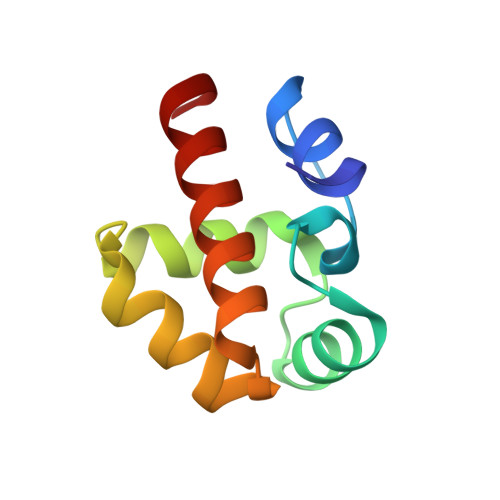
    6RPR

  • PubMed Abstract: 

    Emerin is a nuclear envelope protein that contributes to genome organization and cell mechanics. Through its N-terminal LAP2-emerin-MAN1 (LEM)-domain, emerin interacts with the DNA-binding protein barrier-to-autointegration (BAF). Emerin also binds to members of the linker of the nucleoskeleton and cytoskeleton (LINC) complex. Mutations in the gene encoding emerin are responsible for the majority of cases of X-linked Emery-Dreifuss muscular dystrophy (X-EDMD). Most of these mutations lead to an absence of emerin. A few missense and short deletion mutations in the disordered region of emerin are also associated with X-EDMD. More recently, missense and short deletion mutations P22L, ∆K37 and T43I were discovered in emerin LEM-domain, associated with isolated atrial cardiac defects (ACD). Here we reveal which defects, at both the molecular and cellular levels, are elicited by these LEM-domain mutations. Whereas K37 mutation impaired the correct folding of the LEM-domain, P22L and T43I had no impact on the 3D structure of emerin. Surprisingly, all three mutants bound to BAF, albeit with a weaker affinity in the case of K37. In human myofibroblasts derived from a patient's fibroblasts, emerin ∆K37 was correctly localized at the inner nuclear membrane, but was present at a significantly lower level, indicating that this mutant is abnormally degraded. Moreover, SUN2 was reduced, and these cells were defective in producing actin stress fibers when grown on a stiff substrate and after cyclic stretches. Altogether, our data suggest that the main effect of mutation K37 is to perturb emerin function within the LINC complex in response to mechanical stress.


  • Organizational Affiliation

    Sorbonne Université, INSERM UMR_S974, Center for Research in Myology, 75013 Paris, France. nada.elahmady@gmail.com.


Macromolecules
Find similar proteins by:  (by identity cutoff)  |  3D Structure
Entity ID: 1
MoleculeChains Sequence LengthOrganismDetailsImage
Prelamin-A/CA [auth B]116Homo sapiensMutation(s): 0 
Gene Names: LMNALMN1
UniProt & NIH Common Fund Data Resources
Find proteins for P02545 (Homo sapiens)
Explore P02545 
Go to UniProtKB:  P02545
PHAROS:  P02545
GTEx:  ENSG00000160789 
Entity Groups  
Sequence Clusters30% Identity50% Identity70% Identity90% Identity95% Identity100% Identity
UniProt GroupP02545
Sequence Annotations
Expand
  • Reference Sequence
Find similar proteins by:  (by identity cutoff)  |  3D Structure
Entity ID: 2
MoleculeChains Sequence LengthOrganismDetailsImage
barrier to autointegration factor (BAF)B [auth D],
C [auth E]
87Homo sapiensMutation(s): 0 
UniProt & NIH Common Fund Data Resources
Find proteins for O75531 (Homo sapiens)
Explore O75531 
Go to UniProtKB:  O75531
PHAROS:  O75531
GTEx:  ENSG00000175334 
Entity Groups  
Sequence Clusters30% Identity50% Identity70% Identity90% Identity95% Identity100% Identity
UniProt GroupO75531
Sequence Annotations
Expand
  • Reference Sequence
Find similar proteins by:  (by identity cutoff)  |  3D Structure
Entity ID: 3
MoleculeChains Sequence LengthOrganismDetailsImage
LEM domain of emerin mutant T43ID [auth G]43Homo sapiensMutation(s): 0 
UniProt & NIH Common Fund Data Resources
Find proteins for P50402 (Homo sapiens)
Explore P50402 
Go to UniProtKB:  P50402
PHAROS:  P50402
GTEx:  ENSG00000102119 
Entity Groups  
Sequence Clusters30% Identity50% Identity70% Identity90% Identity95% Identity100% Identity
UniProt GroupP50402
Sequence Annotations
Expand
  • Reference Sequence
Experimental Data & Validation

Experimental Data

  • Method: X-RAY DIFFRACTION
  • Resolution: 2.26 Å
  • R-Value Free: 0.244 
  • R-Value Work: 0.196 
  • R-Value Observed: 0.198 
  • Space Group: C 1 2 1
Unit Cell:
Length ( Å )Angle ( ˚ )
a = 80.2α = 90
b = 81.49β = 121.37
c = 64.88γ = 90
Software Package:
Software NamePurpose
XDSdata reduction
Aimlessdata scaling
BUSTERrefinement
PDB_EXTRACTdata extraction
PHASERphasing

Structure Validation

View Full Validation Report


Ligand Structure Quality Assessment 


Entry History & Funding Information

Deposition Data


Funding OrganizationLocationGrant Number
French Infrastructure for Integrated Structural BiologyFranceANR-10-INSB-05-01
European Communitys Seventh Framework Programme653706

Revision History  (Full details and data files)

  • Version 1.0: 2020-03-04
    Type: Initial release
  • Version 1.1: 2024-01-24
    Changes: Data collection, Database references, Refinement description