6RJR

Crystal structure of a Fungal Catalase at 1.9 Angstrom


Experimental Data Snapshot

  • Method: X-RAY DIFFRACTION
  • Resolution: 1.90 Å
  • R-Value Free: 0.241 
  • R-Value Work: 0.181 
  • R-Value Observed: 0.184 

wwPDB Validation   3D Report Full Report


Ligand Structure Quality Assessment 


This is version 1.1 of the entry. See complete history


Literature

Peroxisomal catalases from the yeasts Pichia pastoris and Kluyveromyces lactis as models for oxidative damage in higher eukaryotes.

Gomez, S.Navas-Yuste, S.Payne, A.M.Rivera, W.Lopez-Estepa, M.Brangbour, C.Fulla, D.Juanhuix, J.Fernandez, F.J.Vega, M.C.

(2019) Free Radic Biol Med 141: 279-290

  • DOI: https://doi.org/10.1016/j.freeradbiomed.2019.06.025
  • Primary Citation of Related Structures:  
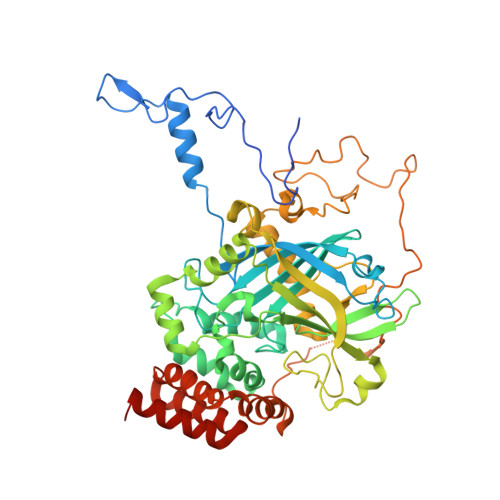
    6RJN, 6RJR

  • PubMed Abstract: 

    Catalases are among the main scavengers of reactive oxygen species (ROS) present in the peroxisome, thereby preventing oxidative cellular and tissular damage. In human, multiple diseases are associated with malfunction of these organelles, which causes accumulation of ROS species and consequently the inefficient detoxification of cells. Despite intense research, much remains to be clarified about the precise molecular role of catalase in cellular homeostasis. Yeast peroxisomes and their peroxisomal catalases have been used as eukaryotic models for oxidative metabolism, ROS generation and detoxification, and associated pathologies. In order to provide reliable models for oxidative metabolism research, we have determined the high-resolution crystal structures of peroxisomal catalase from two important biotechnology and basic biology yeast models, Pichia pastoris and Kluyveromyces lactis. We have performed an extensive functional, biochemical and stability characterization of both enzymes in order to establish their differential activity profiles. Furthermore, we have analyzed the role of the peroxisomal catalase under study in the survival of yeast to oxidative burst challenges combining methanol, water peroxide, and sodium chloride. Interestingly, whereas catalase activity was induced 200-fold upon challenging the methylotrophic P. pastoris cells with methanol, the increase in catalase activity in the non-methylotrophic K. lactis was only moderate. The inhibitory effect of sodium azide and β-mercaptoethanol over both catalases was analyzed, establishing IC50 values for both compounds that are consistent with an elevated resistance of both enzymes toward these inhibitors. Structural comparison of these two novel catalase structures allows us to rationalize the differential susceptibility to inhibitors and oxidative bursts. The inherent worth and validity of the P. pastoris and K. lactis yeast models for oxidative damage will be strengthened by the availability of reliable structural-functional information on these enzymes, which are central to our understanding of peroxisomal response toward oxidative stress.


  • Organizational Affiliation

    Structural and Chemical Biology Department, Center for Biological Research (CIB-CSIC), Madrid, Spain.


Macromolecules
Find similar proteins by:  (by identity cutoff)  |  3D Structure
Entity ID: 1
MoleculeChains Sequence LengthOrganismDetailsImage
Catalase
A, B, C, D
537Kluyveromyces lactisMutation(s): 0 
Gene Names: KLLA0_D11660g
EC: 1.11.1.6
UniProt
Find proteins for Q6CR58 (Kluyveromyces lactis (strain ATCC 8585 / CBS 2359 / DSM 70799 / NBRC 1267 / NRRL Y-1140 / WM37))
Explore Q6CR58 
Go to UniProtKB:  Q6CR58
Entity Groups  
Sequence Clusters30% Identity50% Identity70% Identity90% Identity95% Identity100% Identity
UniProt GroupQ6CR58
Sequence Annotations
Expand
  • Reference Sequence
Small Molecules
Ligands 5 Unique
IDChains Name / Formula / InChI Key2D Diagram3D Interactions
NDP (Subject of Investigation/LOI)
Query on NDP

Download Ideal Coordinates CCD File 
F [auth A],
O [auth B],
T [auth C],
Z [auth D]
NADPH DIHYDRO-NICOTINAMIDE-ADENINE-DINUCLEOTIDE PHOSPHATE
C21 H30 N7 O17 P3
ACFIXJIJDZMPPO-NNYOXOHSSA-N
HEM (Subject of Investigation/LOI)
Query on HEM

Download Ideal Coordinates CCD File 
E [auth A],
N [auth B],
S [auth C],
Y [auth D]
PROTOPORPHYRIN IX CONTAINING FE
C34 H32 Fe N4 O4
KABFMIBPWCXCRK-RGGAHWMASA-L
GOL
Query on GOL

Download Ideal Coordinates CCD File 
AA [auth D]
BA [auth D]
G [auth A]
H [auth A]
I [auth A]
AA [auth D],
BA [auth D],
G [auth A],
H [auth A],
I [auth A],
J [auth A],
K [auth A],
L [auth A],
P [auth B],
U [auth C],
V [auth C]
GLYCEROL
C3 H8 O3
PEDCQBHIVMGVHV-UHFFFAOYSA-N
K
Query on K

Download Ideal Coordinates CCD File 
CA [auth D],
M [auth A],
Q [auth B],
W [auth C]
POTASSIUM ION
K
NPYPAHLBTDXSSS-UHFFFAOYSA-N
CL
Query on CL

Download Ideal Coordinates CCD File 
R [auth B],
X [auth D]
CHLORIDE ION
Cl
VEXZGXHMUGYJMC-UHFFFAOYSA-M
Experimental Data & Validation

Experimental Data

  • Method: X-RAY DIFFRACTION
  • Resolution: 1.90 Å
  • R-Value Free: 0.241 
  • R-Value Work: 0.181 
  • R-Value Observed: 0.184 
  • Space Group: P 21 21 21
Unit Cell:
Length ( Å )Angle ( ˚ )
a = 96.67α = 90
b = 131.62β = 90
c = 176.67γ = 90
Software Package:
Software NamePurpose
PHENIXrefinement
XDSdata reduction
Aimlessdata scaling
PHASERphasing

Structure Validation

View Full Validation Report


Ligand Structure Quality Assessment 


Entry History & Funding Information

Deposition Data


Funding OrganizationLocationGrant Number
Spanish Ministry of Economy and CompetitivenessSpainCTQ2015-66206-C2-2-R
Spanish Ministry of Economy and CompetitivenessSpainSAF2015-72961-EXP
Spanish National Research CouncilSpainPIE-20160E064

Revision History  (Full details and data files)

  • Version 1.0: 2020-03-11
    Type: Initial release
  • Version 1.1: 2024-01-24
    Changes: Data collection, Database references, Derived calculations, Refinement description