6R2M

Crystal structure of PssZ from Listeria monocytogenes


Experimental Data Snapshot

  • Method: X-RAY DIFFRACTION
  • Resolution: 1.62 Å
  • R-Value Free: 0.200 
  • R-Value Work: 0.169 
  • R-Value Observed: 0.171 

wwPDB Validation   3D Report Full Report


This is version 1.1 of the entry. See complete history


Literature

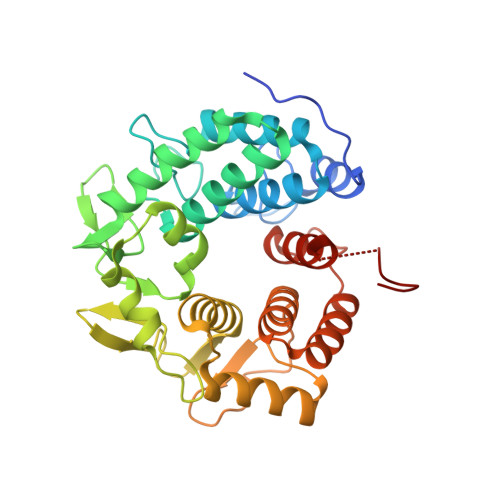
Crystal structure of the glycoside hydrolase PssZ from Listeria monocytogenes.

Wu, H.Qiao, S.Li, D.Guo, L.Zhu, M.Ma, L.Z.

(2019) Acta Crystallogr F Struct Biol Commun 75: 501-506

  • DOI: https://doi.org/10.1107/S2053230X19008100
  • Primary Citation of Related Structures:  
    6R2M

  • PubMed Abstract: 

    Biofilms are microbial communities that are embedded in the extracellular matrix. The exopolysaccharide (EPS) is a key component of the biofilm matrix that maintains the structure of the biofilm and protects the bacteria from antimicrobials. Microbial glycoside hydrolases have been exploited to disrupt biofilms by breaking down EPSs. PssZ has recently been identified as a glycoside hydrolase that can disperse aggregates of Listeria monocytogenes. In this study, the crystal structure of PssZ has been determined at 1.6 Å resolution. PssZ belongs to glycoside hydrolase family 8 and adopts a classical (α/α) 6 -barrel fold. This architecture forms a deep groove which may serve as the substrate-binding pocket. The conserved catalytic residues (Glu72, Trp110, Asn119, Phe167, Tyr183 and Asp232) are localized at the centre of the groove. This crystal structure will help to improve the understanding of the hydrolytic mechanism of PssZ and its application as a biofilm disrupter.


  • Organizational Affiliation

    State Key Laboratory of Microbial Resources, Institute of Microbiology, Chinese Academy of Sciences, Beijing 100101, People's Republic of China.


Macromolecules
Find similar proteins by:  (by identity cutoff)  |  3D Structure
Entity ID: 1
MoleculeChains Sequence LengthOrganismDetailsImage
Glycoside transferase
A, B
326Listeria monocytogenesMutation(s): 0 
Gene Names: RK57_03105
UniProt
Find proteins for A0A3D7VE02 (Listeria monocytogenes)
Explore A0A3D7VE02 
Go to UniProtKB:  A0A3D7VE02
Entity Groups  
Sequence Clusters30% Identity50% Identity70% Identity90% Identity95% Identity100% Identity
UniProt GroupA0A3D7VE02
Sequence Annotations
Expand
  • Reference Sequence
Experimental Data & Validation

Experimental Data

  • Method: X-RAY DIFFRACTION
  • Resolution: 1.62 Å
  • R-Value Free: 0.200 
  • R-Value Work: 0.169 
  • R-Value Observed: 0.171 
  • Space Group: P 1 21 1
Unit Cell:
Length ( Å )Angle ( ˚ )
a = 40.08α = 90
b = 71.56β = 95.8
c = 131.3γ = 90
Software Package:
Software NamePurpose
PHENIXrefinement
XDSdata reduction
XSCALEdata scaling
PHENIXphasing

Structure Validation

View Full Validation Report


Entry History 

Deposition Data

Revision History  (Full details and data files)

  • Version 1.0: 2019-07-24
    Type: Initial release
  • Version 1.1: 2019-09-25
    Changes: Data collection, Database references, Source and taxonomy