6QXL

Crystal Structure of Pyruvate Kinase II (PykA) from Pseudomonas aeruginosa in complex with sodium malonate, magnesium and glucose-6-phosphate


Experimental Data Snapshot

  • Method: X-RAY DIFFRACTION
  • Resolution: 2.43 Å
  • R-Value Free: 0.251 
  • R-Value Work: 0.227 
  • R-Value Observed: 0.228 

wwPDB Validation   3D Report Full Report


Ligand Structure Quality Assessment 


This is version 1.4 of the entry. See complete history


Literature

Evolutionary plasticity in the allosteric regulator-binding site of pyruvate kinase isoform PykA fromPseudomonas aeruginosa.

Abdelhamid, Y.Brear, P.Greenhalgh, J.Chee, X.Rahman, T.Welch, M.

(2019) J Biol Chem 294: 15505-15516

  • DOI: https://doi.org/10.1074/jbc.RA119.009156
  • Primary Citation of Related Structures:  
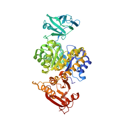
    6QXL

  • PubMed Abstract: 

    Unlike many other well-characterized bacteria, the opportunistic human pathogen Pseudomonas aeruginosa relies exclusively on the Entner-Doudoroff pathway (EDP) for glycolysis. Pyruvate kinase (PK) is the main "pacemaker" of the EDP, and its activity is also relevant for P. aeruginosa virulence. Two distinct isozymes of bacterial PK have been recognized, PykA and PykF. Here, using growth and expression analyses of relevant PK mutants, we show that PykA is the dominant isoform in P. aeruginosa Enzyme kinetics assays revealed that PykA displays potent K-type allosteric activation by glucose 6-phosphate and by intermediates from the pentose phosphate pathway. Unexpectedly, the X-ray structure of PykA at 2.4 Å resolution revealed that glucose 6-phosphate binds in a pocket that is distinct from the binding site reported for this metabolite in the PK from Mycobacterium tuberculosis (the only other available bacterial PK structure containing bound glucose 6-phosphate). We propose a mechanism by which glucose 6-phosphate binding at the allosteric site communicates with the PykA active site. Taken together, our findings indicate remarkable evolutionary plasticity in the mechanism(s) by which PK senses and responds to allosteric signals.


  • Organizational Affiliation

    Department of Biochemistry, University of Cambridge, Cambridge CB2 1QW, United Kingdom.


Macromolecules
Find similar proteins by:  (by identity cutoff)  |  3D Structure
Entity ID: 1
MoleculeChains Sequence LengthOrganismDetailsImage
Pyruvate kinase
A, B, C, D, E
A, B, C, D, E, F, G, H, I, J, K, L
483Pseudomonas aeruginosaMutation(s): 0 
Gene Names: pykApykpykA_1C8257_26620CAZ10_21690DZ940_08845NCTC13719_04679PAERUG_E15_London_28_01_14_03018PAMH19_3832RW109_RW109_05666
EC: 2.7.1.40
UniProt
Find proteins for Q9HW72 (Pseudomonas aeruginosa (strain ATCC 15692 / DSM 22644 / CIP 104116 / JCM 14847 / LMG 12228 / 1C / PRS 101 / PAO1))
Explore Q9HW72 
Go to UniProtKB:  Q9HW72
Entity Groups  
Sequence Clusters30% Identity50% Identity70% Identity90% Identity95% Identity100% Identity
UniProt GroupQ9HW72
Sequence Annotations
Expand
  • Reference Sequence
Small Molecules
Ligands 4 Unique
IDChains Name / Formula / InChI Key2D Diagram3D Interactions
G6P (Subject of Investigation/LOI)
Query on G6P

Download Ideal Coordinates CCD File 
AA [auth E]
EA [auth F]
HA [auth G]
KA [auth H]
M [auth A]
AA [auth E],
EA [auth F],
HA [auth G],
KA [auth H],
M [auth A],
NA [auth I],
P [auth B],
QA [auth J],
T [auth C],
TA [auth K],
W [auth D],
WA [auth L]
6-O-phosphono-alpha-D-glucopyranose
C6 H13 O9 P
NBSCHQHZLSJFNQ-DVKNGEFBSA-N
MLI (Subject of Investigation/LOI)
Query on MLI

Download Ideal Coordinates CCD File 
DA [auth E]
GA [auth F]
JA [auth G]
MA [auth H]
O [auth A]
DA [auth E],
GA [auth F],
JA [auth G],
MA [auth H],
O [auth A],
PA [auth I],
S [auth B],
SA [auth J],
V [auth C],
VA [auth K],
YA [auth L],
Z [auth D]
MALONATE ION
C3 H2 O4
OFOBLEOULBTSOW-UHFFFAOYSA-L
GOL
Query on GOL

Download Ideal Coordinates CCD File 
CA [auth E],
R [auth B],
Y [auth D]
GLYCEROL
C3 H8 O3
PEDCQBHIVMGVHV-UHFFFAOYSA-N
MG (Subject of Investigation/LOI)
Query on MG

Download Ideal Coordinates CCD File 
BA [auth E]
FA [auth F]
IA [auth G]
LA [auth H]
N [auth A]
BA [auth E],
FA [auth F],
IA [auth G],
LA [auth H],
N [auth A],
OA [auth I],
Q [auth B],
RA [auth J],
U [auth C],
UA [auth K],
X [auth D],
XA [auth L]
MAGNESIUM ION
Mg
JLVVSXFLKOJNIY-UHFFFAOYSA-N
Experimental Data & Validation

Experimental Data

  • Method: X-RAY DIFFRACTION
  • Resolution: 2.43 Å
  • R-Value Free: 0.251 
  • R-Value Work: 0.227 
  • R-Value Observed: 0.228 
  • Space Group: P 31 2 1
Unit Cell:
Length ( Å )Angle ( ˚ )
a = 182.477α = 90
b = 182.477β = 90
c = 405.044γ = 120
Software Package:
Software NamePurpose
BUSTERrefinement
XDSdata reduction
Aimlessdata scaling
PHASERphasing
PDB_EXTRACTdata extraction

Structure Validation

View Full Validation Report


Ligand Structure Quality Assessment 


Entry History & Funding Information

Deposition Data


Funding OrganizationLocationGrant Number
Yousef Jameel ScholarshipUnited Kingdom--

Revision History  (Full details and data files)

  • Version 1.0: 2019-09-11
    Type: Initial release
  • Version 1.1: 2019-09-18
    Changes: Data collection, Database references
  • Version 1.2: 2019-10-30
    Changes: Data collection, Database references
  • Version 1.3: 2020-07-29
    Type: Remediation
    Reason: Carbohydrate remediation
    Changes: Data collection, Derived calculations, Structure summary
  • Version 1.4: 2024-01-24
    Changes: Data collection, Database references, Refinement description, Structure summary