6QKY

Tryptophan synthase subunit alpha from Streptococcus pneumoniae with 3D domain swap in the core of TIM barrel


Experimental Data Snapshot

  • Method: X-RAY DIFFRACTION
  • Resolution: 2.54 Å
  • R-Value Free: 0.234 
  • R-Value Work: 0.193 
  • R-Value Observed: 0.194 

wwPDB Validation   3D Report Full Report


This is version 1.2 of the entry. See complete history


Literature

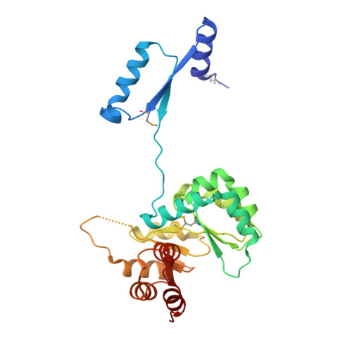
3D domain swapping in the TIM barrel of the alpha subunit of Streptococcus pneumoniae tryptophan synthase.

Michalska, K.Kowiel, M.Bigelow, L.Endres, M.Gilski, M.Jaskolski, M.Joachimiak, A.

(2020) Acta Crystallogr D Struct Biol 76: 166-175

  • DOI: https://doi.org/10.1107/S2059798320000212
  • Primary Citation of Related Structures:  
    6QKY

  • PubMed Abstract: 

    Tryptophan synthase catalyzes the last two steps of tryptophan biosynthesis in plants, fungi and bacteria. It consists of two protein chains, designated α and β, encoded by trpA and trpB genes, that function as an αββα complex. Structural and functional features of tryptophan synthase have been extensively studied, explaining the roles of individual residues in the two active sites in catalysis and allosteric regulation. TrpA serves as a model for protein-folding studies. In 1969, Jackson and Yanofsky observed that the typically monomeric TrpA forms a small population of dimers. Dimerization was postulated to take place through an exchange of structural elements of the monomeric chains, a phenomenon later termed 3D domain swapping. The structural details of the TrpA dimer have remained unknown. Here, the crystal structure of the Streptococcus pneumoniae TrpA homodimer is reported, demonstrating 3D domain swapping in a TIM-barrel fold for the first time. The N-terminal domain comprising the H0-S1-H1-S2 elements is exchanged, while the hinge region corresponds to loop L2 linking strand S2 to helix H2'. The structural elements S2 and L2 carry the catalytic residues Glu52 and Asp63. As the S2 element is part of the swapped domain, the architecture of the catalytic apparatus in the dimer is recreated from two protein chains. The homodimer interface overlaps with the α-β interface of the tryptophan synthase αββα heterotetramer, suggesting that the 3D domain-swapped dimer cannot form a complex with the β subunit. In the crystal, the dimers assemble into a decamer comprising two pentameric rings.


  • Organizational Affiliation

    Midwest Center for Structural Genomics, X-ray Science Division, Argonne National Laboratory, Argonne, IL 60439, USA.


Macromolecules
Find similar proteins by:  (by identity cutoff)  |  3D Structure
Entity ID: 1
MoleculeChains Sequence LengthOrganismDetailsImage
Tryptophan synthase alpha chain
A, B, C, D, E
A, B, C, D, E, F, G, H, I, J
261Streptococcus pneumoniaeMutation(s): 0 
Gene Names: trpASP_1811
EC: 4.2.1.20
UniProt
Find proteins for Q97P33 (Streptococcus pneumoniae serotype 4 (strain ATCC BAA-334 / TIGR4))
Explore Q97P33 
Go to UniProtKB:  Q97P33
Entity Groups  
Sequence Clusters30% Identity50% Identity70% Identity90% Identity95% Identity100% Identity
UniProt GroupQ97P33
Sequence Annotations
Expand
  • Reference Sequence
Small Molecules
Ligands 3 Unique
IDChains Name / Formula / InChI Key2D Diagram3D Interactions
PEG
Query on PEG

Download Ideal Coordinates CCD File 
M [auth B],
S [auth E]
DI(HYDROXYETHYL)ETHER
C4 H10 O3
MTHSVFCYNBDYFN-UHFFFAOYSA-N
GOL
Query on GOL

Download Ideal Coordinates CCD File 
K [auth A],
O [auth C],
P [auth C]
GLYCEROL
C3 H8 O3
PEDCQBHIVMGVHV-UHFFFAOYSA-N
ACY
Query on ACY

Download Ideal Coordinates CCD File 
L [auth A]
N [auth B]
Q [auth C]
R [auth D]
T [auth E]
L [auth A],
N [auth B],
Q [auth C],
R [auth D],
T [auth E],
U [auth E],
V [auth G],
W [auth H],
X [auth I],
Y [auth J]
ACETIC ACID
C2 H4 O2
QTBSBXVTEAMEQO-UHFFFAOYSA-N
Modified Residues  1 Unique
IDChains TypeFormula2D DiagramParent
MSE
Query on MSE
A, B, C, D, E
A, B, C, D, E, F, G, H, I, J
L-PEPTIDE LINKINGC5 H11 N O2 SeMET
Experimental Data & Validation

Experimental Data

Unit Cell:
Length ( Å )Angle ( ˚ )
a = 104.81α = 90
b = 138.62β = 117.21
c = 107.25γ = 90
Software Package:
Software NamePurpose
REFMACrefinement
XDSdata reduction
XSCALEdata scaling
CRANK2phasing
SBC-Collectdata collection

Structure Validation

View Full Validation Report


Entry History & Funding Information

Deposition Data


Funding OrganizationLocationGrant Number
National Institutes of Health/National Institute Of Allergy and Infectious Diseases (NIH/NIAID)United StatesHHSN272201700060C

Revision History  (Full details and data files)

  • Version 1.0: 2019-03-27
    Type: Initial release
  • Version 1.1: 2020-02-19
    Changes: Database references
  • Version 1.2: 2022-03-30
    Changes: Author supporting evidence, Database references