6QAP

Structure of the human aldehyde dehydrogenase 9A1 in C2 space group


Experimental Data Snapshot

  • Method: X-RAY DIFFRACTION
  • Resolution: 2.30 Å
  • R-Value Free: 0.251 
  • R-Value Work: 0.220 
  • R-Value Observed: 0.222 

wwPDB Validation   3D Report Full Report


This is version 1.2 of the entry. See complete history


Literature

Kinetic and structural analysis of human ALDH9A1.

Koncitikova, R.Vigouroux, A.Kopecna, M.Sebela, M.Morera, S.Kopecny, D.

(2019) Biosci Rep 39

  • DOI: https://doi.org/10.1042/BSR20190558
  • Primary Citation of Related Structures:  
    6QAK, 6QAO, 6QAP

  • PubMed Abstract: 

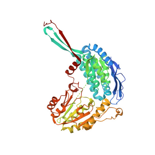
    Aldehyde dehydrogenases (ALDHs) constitute a superfamily of NAD(P) + -dependent enzymes, which detoxify aldehydes produced in various metabolic pathways to the corresponding carboxylic acids. Among the 19 human ALDHs, the cytosolic ALDH9A1 has so far never been fully enzymatically characterized and its structure is still unknown. Here, we report complete molecular and kinetic properties of human ALDH9A1 as well as three crystal forms at 2.3, 2.9, and 2.5 Å resolution. We show that ALDH9A1 exhibits wide substrate specificity to aminoaldehydes, aliphatic and aromatic aldehydes with a clear preference for γ -trimethylaminobutyraldehyde (TMABAL). The structure of ALDH9A1 reveals that the enzyme assembles as a tetramer. Each ALDH monomer displays a typical ALDHs fold composed of an oligomerization domain, a coenzyme domain, a catalytic domain, and an inter-domain linker highly conserved in amino-acid sequence and folding. Nonetheless, structural comparison reveals a position and a fold of the inter-domain linker of ALDH9A1 never observed in any other ALDH so far. This unique difference is not compatible with the presence of a bound substrate and a large conformational rearrangement of the linker up to 30 Å has to occur to allow the access of the substrate channel. Moreover, the αβE region consisting of an α-helix and a β-strand of the coenzyme domain at the dimer interface are disordered, likely due to the loss of interactions with the inter-domain linker, which leads to incomplete β-nicotinamide adenine dinucleotide (NAD + ) binding pocket.


  • Organizational Affiliation

    Department of Protein Biochemistry and Proteomics, Centre of the Region Haná for Biotechnological and Agricultural Research, Faculty of Science, Palacký University, Šlechtitelů 27, CZ-783 71 Olomouc, Czech Republic.


Macromolecules
Find similar proteins by:  (by identity cutoff)  |  3D Structure
Entity ID: 1
MoleculeChains Sequence LengthOrganismDetailsImage
4-trimethylaminobutyraldehyde dehydrogenase
A, B, C, D
508Homo sapiensMutation(s): 0 
Gene Names: ALDH9A1ALDH4ALDH7ALDH9
EC: 1.2.1.47 (PDB Primary Data), 1.2.1.3 (PDB Primary Data), 1.2.1.19 (PDB Primary Data)
UniProt & NIH Common Fund Data Resources
Find proteins for P49189 (Homo sapiens)
Explore P49189 
Go to UniProtKB:  P49189
PHAROS:  P49189
GTEx:  ENSG00000143149 
Entity Groups  
Sequence Clusters30% Identity50% Identity70% Identity90% Identity95% Identity100% Identity
UniProt GroupP49189
Sequence Annotations
Expand
  • Reference Sequence
Small Molecules
Ligands 3 Unique
IDChains Name / Formula / InChI Key2D Diagram3D Interactions
PEG
Query on PEG

Download Ideal Coordinates CCD File 
M [auth B],
R [auth C],
S [auth C],
T [auth C]
DI(HYDROXYETHYL)ETHER
C4 H10 O3
MTHSVFCYNBDYFN-UHFFFAOYSA-N
EDO
Query on EDO

Download Ideal Coordinates CCD File 
AA [auth D]
BA [auth D]
CA [auth D]
DA [auth D]
E [auth A]
AA [auth D],
BA [auth D],
CA [auth D],
DA [auth D],
E [auth A],
EA [auth D],
F [auth A],
G [auth A],
H [auth A],
I [auth A],
J [auth B],
K [auth B],
L [auth B],
Q [auth C],
Z [auth D]
1,2-ETHANEDIOL
C2 H6 O2
LYCAIKOWRPUZTN-UHFFFAOYSA-N
NA
Query on NA

Download Ideal Coordinates CCD File 
N [auth C]
O [auth C]
P [auth C]
U [auth D]
V [auth D]
N [auth C],
O [auth C],
P [auth C],
U [auth D],
V [auth D],
W [auth D],
X [auth D],
Y [auth D]
SODIUM ION
Na
FKNQFGJONOIPTF-UHFFFAOYSA-N
Experimental Data & Validation

Experimental Data

  • Method: X-RAY DIFFRACTION
  • Resolution: 2.30 Å
  • R-Value Free: 0.251 
  • R-Value Work: 0.220 
  • R-Value Observed: 0.222 
  • Space Group: C 1 2 1
Unit Cell:
Length ( Å )Angle ( ˚ )
a = 164.29α = 90
b = 160.03β = 91.09
c = 84.6γ = 90
Software Package:
Software NamePurpose
BUSTERrefinement
XDSdata reduction
XSCALEdata scaling
PHASERphasing

Structure Validation

View Full Validation Report


Entry History 

Deposition Data

Revision History  (Full details and data files)

  • Version 1.0: 2019-04-10
    Type: Initial release
  • Version 1.1: 2019-05-08
    Changes: Data collection, Database references
  • Version 1.2: 2024-01-24
    Changes: Data collection, Database references, Derived calculations, Refinement description