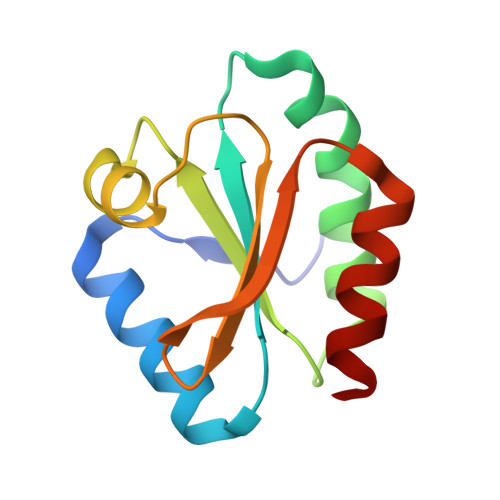
Structural and Biochemical Insights into the Reactivity of Thioredoxin h1 fromChlamydomonas reinhardtii.
Marchand, C.H., Fermani, S., Rossi, J., Gurrieri, L., Tedesco, D., Henri, J., Sparla, F., Trost, P., Lemaire, S.D., Zaffagnini, M.(2019) Antioxidants (Basel) 8
- PubMed: 30609656
- DOI: https://doi.org/10.3390/antiox8010010
- Primary Citation of Related Structures:
6Q46, 6Q47, 6Q6T, 6Q6U, 6Q6V - PubMed Abstract:
Thioredoxins (TRXs) are major protein disulfide reductases of the cell. Their redox activity relies on a conserved Trp-Cys-(Gly/Pro)-Pro-Cys active site bearing two cysteine (Cys) residues that can be found either as free thiols (reduced TRXs) or linked together by a disulfide bond (oxidized TRXs) during the catalytic cycle. Their reactivity is crucial for TRX activity, and depends on the active site microenvironment. Here, we solved and compared the 3D structure of reduced and oxidized TRX h1 from Chlamydomonas reinhardtii (CrTRXh1). The three-dimensional structure was also determined for mutants of each active site Cys. Structural alignments of CrTRXh1 with other structurally solved plant TRXs showed a common spatial fold, despite the low sequence identity. Structural analyses of CrTRXh1 revealed that the protein adopts an identical conformation independently from its redox state. Treatment with iodoacetamide (IAM), a Cys alkylating agent, resulted in a rapid and pH-dependent inactivation of CrTRXh1. Starting from fully reduced CrTRXh1, we determined the acid dissociation constant (p K a ) of each active site Cys by Matrix-assisted laser desorption/ionization-time of flight (MALDI-TOF) mass spectrometry analyses coupled to differential IAM-based alkylation. Based on the diversity of catalytic Cys deprotonation states, the mechanisms and structural features underlying disulfide redox activity are discussed.
Organizational Affiliation:
Laboratoire de Biologie Moléculaire et Cellulaire des Eucaryotes, Institut de Biologie Physico-Chimique, Unité Mixte de Recherche 8226 CNRS Sorbonne Université, 13 rue Pierre et Marie Curie, 75005 Paris, France. christophe.marchand@ibpc.fr.