MicroED with the Falcon III direct electron detector.
Hattne, J., Martynowycz, M.W., Penczek, P.A., Gonen, T.(2019) IUCrJ 6: 921-926
- PubMed: 31576224
- DOI: https://doi.org/10.1107/S2052252519010583
- Primary Citation of Related Structures:
6PU4, 6PU5 - PubMed Abstract:
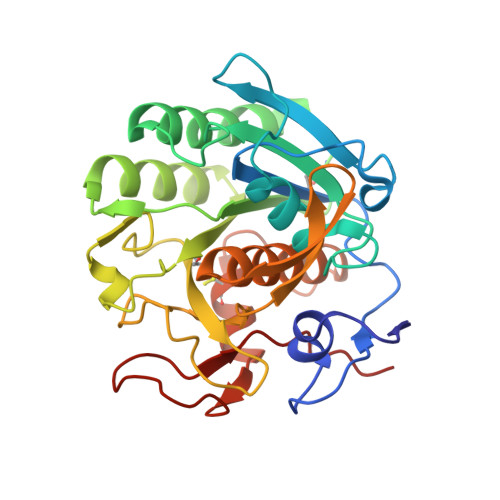
Microcrystal electron diffraction (MicroED) combines crystallography and electron cryo-microscopy (cryo-EM) into a method that is applicable to high-resolution structure determination. In MicroED, nanosized crystals, which are often intractable using other techniques, are probed by high-energy electrons in a transmission electron microscope. Diffraction data are recorded by a camera in movie mode: the nanocrystal is continuously rotated in the beam, thus creating a sequence of frames that constitute a movie with respect to the rotation angle. Until now, diffraction-optimized cameras have mostly been used for MicroED. Here, the use of a direct electron detector that was designed for imaging is reported. It is demonstrated that data can be collected more rapidly using the Falcon III for MicroED and with markedly lower exposure than has previously been reported. The Falcon III was operated at 40 frames per second and complete data sets reaching atomic resolution were recorded in minutes. The resulting density maps to 2.1 Å resolution of the serine protease proteinase K showed no visible signs of radiation damage. It is thus demonstrated that dedicated diffraction-optimized detectors are not required for MicroED, as shown by the fact that the very same cameras that are used for imaging applications in electron microscopy, such as single-particle cryo-EM, can also be used effectively for diffraction measurements.
Organizational Affiliation:
Howard Hughes Medical Institute, Department of Biological Chemistry, David Geffen School of Medicine, University of California, Los Angeles, CA 90095, USA.