Functionally critical residues in the aminoglycoside resistance-associated methyltransferase RmtC play distinct roles in 30S substrate recognition.
Nosrati, M., Dey, D., Mehrani, A., Strassler, S.E., Zelinskaya, N., Hoffer, E.D., Stagg, S.M., Dunham, C.M., Conn, G.L.(2019) J Biol Chem 294: 17642-17653
- PubMed: 31594862
- DOI: https://doi.org/10.1074/jbc.RA119.011181
- Primary Citation of Related Structures:
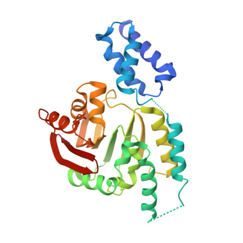
6PQB - PubMed Abstract:
Methylation of the small ribosome subunit rRNA in the ribosomal decoding center results in exceptionally high-level aminoglycoside resistance in bacteria. Enzymes that methylate 16S rRNA on N7 of nucleotide G1405 (m 7 G1405) have been identified in both aminoglycoside-producing and clinically drug-resistant pathogenic bacteria. Using a fluorescence polarization 30S-binding assay and a new crystal structure of the methyltransferase RmtC at 3.14 Å resolution, here we report a structure-guided functional study of 30S substrate recognition by the aminoglycoside resistance-associated 16S rRNA (m 7 G1405) methyltransferases. We found that the binding site for these enzymes in the 30S subunit directly overlaps with that of a second family of aminoglycoside resistance-associated 16S rRNA (m 1 A1408) methyltransferases, suggesting that both groups of enzymes may exploit the same conserved rRNA tertiary surface for docking to the 30S. Within RmtC, we defined an N-terminal domain surface, comprising basic residues from both the N1 and N2 subdomains, that directly contributes to 30S-binding affinity. In contrast, additional residues lining a contiguous adjacent surface on the C-terminal domain were critical for 16S rRNA modification but did not directly contribute to the binding affinity. The results from our experiments define the critical features of m 7 G1405 methyltransferase-substrate recognition and distinguish at least two distinct, functionally critical contributions of the tested enzyme residues: 30S-binding affinity and stabilizing a binding-induced 16S rRNA conformation necessary for G1405 modification. Our study sets the scene for future high-resolution structural studies of the 30S-methyltransferase complex and for potential exploitation of unique aspects of substrate recognition in future therapeutic strategies.
Organizational Affiliation:
Department of Biochemistry, Emory University School of Medicine, Atlanta, Georgia 30322.