6PI7

Crystal structure of the TDRD2 extended Tudor domain in complex with an antibody fragment and the PIWIL1 peptide


Experimental Data Snapshot

  • Method: X-RAY DIFFRACTION
  • Resolution: 2.80 Å
  • R-Value Free: 0.206 
  • R-Value Work: 0.190 
  • R-Value Observed: 0.191 

wwPDB Validation   3D Report Full Report


This is version 1.2 of the entry. See complete history


Literature

Lesson from a Fab-enabled co-crystallization study of TDRD2 and PIWIL1.

Chen, S.Zhang, W.Min, J.Liu, K.

(2020) Methods 175: 72-78

  • DOI: https://doi.org/10.1016/j.ymeth.2019.07.002
  • Primary Citation of Related Structures:  
    6PI7

  • PubMed Abstract: 

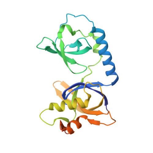
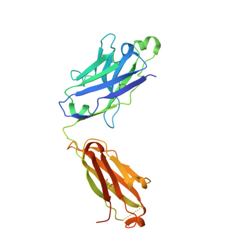
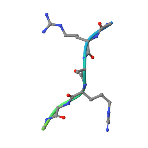
    The interaction of Tudor domain-containing proteins (TDRDs) with P-element-induced wimpy testis (PIWI) proteins plays critical roles in transposon silencing and spermatogenesis. Most human TDRDs recognize PIWI proteins in a methylarginine-dependent manner via their extended Tudor (eTudor) domains, except TDRD2, which prefers an unmethylated PIWI protein. In order to illustrate the recognition of unmethylated PIWI proteins by TDRD2, we extensively tried co-crystallization of the TDRD2 eTudor with different PIWIL1 peptides, but to no avail. Recombinant antigen-binding fragments (Fabs) have been used to crystallize some difficult proteins in the past, so we generated Fab against the TDRD2 eTudor protein using a phage-display antibody library, and one of these Fab fragments indeed facilitated the co-crystallization of TDRD2 and PIWIL1. Structural analysis of Fab, the TDRD2 eTudor domain in complex with an unmethylated PIWIL1-derived peptide revealed that the PIWIL1 residues G3 through R8 bound between the Tudor core and SN domain of TDRD2. The C-terminal residues of the PIWIL1 peptide were not resolved, presumably due to steric competition with the heavy chain of the Fab. We propose Fab-assisted crystallization as a tool not only for structural studies of single proteins, but also for analysis of interactions between proteins and their ligands in cases where co-crystallization of native protein complexes fails.


  • Organizational Affiliation

    Hubei Key Laboratory of Genetic Regulation and Integrative Biology, School of Life Sciences, Central China Normal University, Wuhan 430079, PR China.


Macromolecules
Find similar proteins by:  (by identity cutoff)  |  3D Structure
Entity ID: 1
MoleculeChains Sequence LengthOrganismDetailsImage
Tudor and KH domain-containing protein
A, D
222Homo sapiensMutation(s): 0 
Gene Names: TDRKHTDRD2
UniProt & NIH Common Fund Data Resources
Find proteins for Q9Y2W6 (Homo sapiens)
Explore Q9Y2W6 
Go to UniProtKB:  Q9Y2W6
PHAROS:  Q9Y2W6
GTEx:  ENSG00000182134 
Entity Groups  
Sequence Clusters30% Identity50% Identity70% Identity90% Identity95% Identity100% Identity
UniProt GroupQ9Y2W6
Sequence Annotations
Expand
  • Reference Sequence
Find similar proteins by:  (by identity cutoff)  |  3D Structure
Entity ID: 2
MoleculeChains Sequence LengthOrganismDetailsImage
Uncharacterized protein
B, E
226Homo sapiensMutation(s): 0 
Entity Groups  
Sequence Clusters30% Identity50% Identity70% Identity90% Identity95% Identity100% Identity
Sequence Annotations
Expand
  • Reference Sequence
Find similar proteins by:  (by identity cutoff)  |  3D Structure
Entity ID: 3
MoleculeChains Sequence LengthOrganismDetailsImage
Fab antigen-binding fragment
C, F
244Homo sapiensMutation(s): 0 
Entity Groups  
Sequence Clusters30% Identity50% Identity70% Identity90% Identity95% Identity100% Identity
Sequence Annotations
Expand
  • Reference Sequence

Find similar proteins by:  Sequence   |   3D Structure  

Entity ID: 4
MoleculeChains Sequence LengthOrganismDetailsImage
Piwi-like protein 116Homo sapiensMutation(s): 0 
UniProt & NIH Common Fund Data Resources
Find proteins for Q96J94 (Homo sapiens)
Explore Q96J94 
Go to UniProtKB:  Q96J94
PHAROS:  Q96J94
GTEx:  ENSG00000125207 
Entity Groups  
Sequence Clusters30% Identity50% Identity70% Identity90% Identity95% Identity100% Identity
UniProt GroupQ96J94
Sequence Annotations
Expand
  • Reference Sequence
Experimental Data & Validation

Experimental Data

  • Method: X-RAY DIFFRACTION
  • Resolution: 2.80 Å
  • R-Value Free: 0.206 
  • R-Value Work: 0.190 
  • R-Value Observed: 0.191 
  • Space Group: P 63
  • Diffraction Data: https://doi.org/10.18430/m36pi7
Unit Cell:
Length ( Å )Angle ( ˚ )
a = 148.099α = 90
b = 148.099β = 90
c = 168.026γ = 120
Software Package:
Software NamePurpose
Aimlessdata scaling
REFMACrefinement
PDB_EXTRACTdata extraction
XDSdata reduction
PHASERphasing
MOLREPphasing

Structure Validation

View Full Validation Report


Entry History 

Deposition Data

Revision History  (Full details and data files)

  • Version 1.0: 2019-07-24
    Type: Initial release
  • Version 1.1: 2020-04-22
    Changes: Database references
  • Version 1.2: 2023-10-11
    Changes: Data collection, Database references, Refinement description