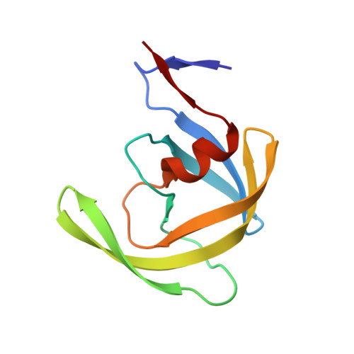
Highly drug-resistant HIV-1 protease reveals decreased intra-subunit interactions due to clusters of mutations.
Kneller, D.W., Agniswamy, J., Harrison, R.W., Weber, I.T.(2020) FEBS J 287: 3235-3254
- PubMed: 31920003
- DOI: https://doi.org/10.1111/febs.15207
- Primary Citation of Related Structures:
6P9A, 6P9B - PubMed Abstract:
Drug-resistance is a serious problem for treatment of the HIV/AIDS pandemic. Potent clinical inhibitors of HIV-1 protease show several orders of magnitude worse inhibition of highly drug-resistant variants. Hence, the structure and enzyme activities were analyzed for HIV protease mutant HIV-1 protease (EC 3.4.23.16) (PR) with 22 mutations (PRS5B) from a clinical isolate that was selected by machine learning to represent high-level drug-resistance. PRS5B has 22 mutations including only one (I84V) in the inhibitor binding site; however, clinical inhibitors had poor inhibition of PRS5B activity with kinetic inhibition value (K i ) values of 4-1000 nm or 18- to 8000-fold worse than for wild-type PR. High-resolution crystal structures of PRS5B complexes with the best inhibitors, amprenavir (APV) and darunavir (DRV) (K i ~ 4 nm), revealed only minor changes in protease-inhibitor interactions. Instead, two distinct clusters of mutations in distal regions induce coordinated conformational changes that decrease favorable internal interactions across the entire protein subunit. The largest structural rearrangements are described and compared to other characterized resistant mutants. In the protease hinge region, the N83D mutation eliminates a hydrogen bond connecting the hinge and core of the protease and increases disorder compared to highly resistant mutants PR with 17 mutations and PR with 20 mutations with similar hinge mutations. In a distal β-sheet, mutations G73T and A71V coordinate with accessory mutations to bring about shifts that propagate throughout the subunit. Molecular dynamics simulations of ligand-free dimers show differences consistent with loss of interactions in mutant compared to wild-type PR. Clusters of mutations exhibit both coordinated and antagonistic effects, suggesting PRS5B may represent an intermediate stage in the evolution of more highly resistant variants. DATABASES: Structural data are available in Protein Data Bank under the accession codes 6P9A and 6P9B for PRS5B/DRV and PRS5B/APV, respectively.
Organizational Affiliation:
Department of Biology, Georgia State University, Atlanta, GA, USA.