6P7E

Structure of T7 DNA Polymerase Bound to a Primer/Template DNA and a Peptide that Mimics the C-terminal Tail of the Primase-Helicase


Experimental Data Snapshot

  • Method: X-RAY DIFFRACTION
  • Resolution: 3.00 Å
  • R-Value Free: 0.248 
  • R-Value Work: 0.214 
  • R-Value Observed: 0.216 

wwPDB Validation   3D Report Full Report


Ligand Structure Quality Assessment 


This is version 1.1 of the entry. See complete history


Literature

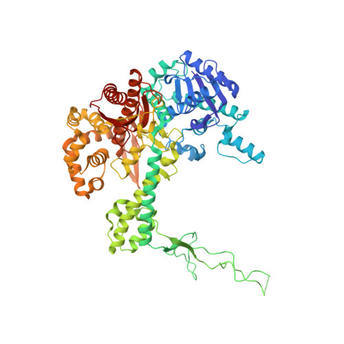
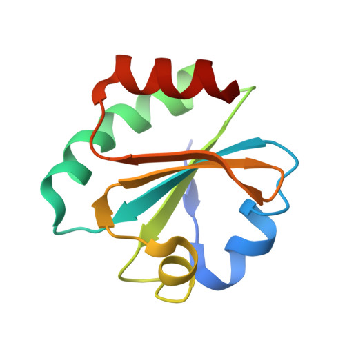
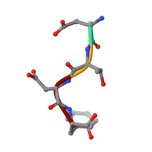
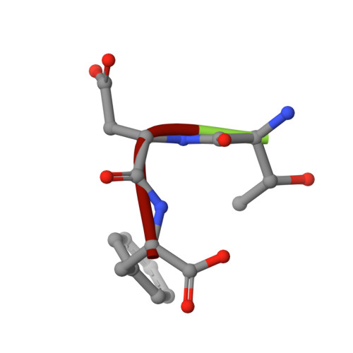
Combined Solution and Crystal Methods Reveal the Electrostatic Tethers That Provide a Flexible Platform for Replication Activities in the Bacteriophage T7 Replisome.

Foster, B.M.Rosenberg, D.Salvo, H.Stephens, K.L.Bintz, B.J.Hammel, M.Ellenberger, T.Gainey, M.D.Wallen, J.R.

(2019) Biochemistry 58: 4466-4479

  • DOI: https://doi.org/10.1021/acs.biochem.9b00525
  • Primary Citation of Related Structures:  
    6P7E

  • PubMed Abstract: 

    Recent structural studies of the bacteriophage T7 DNA replication system have shed light on how multiple proteins assemble to copy two antiparallel DNA strands. In T7, acidic C-terminal tails of both the primase-helicase and single-stranded DNA binding protein bind to two basic patches on the DNA polymerase to aid in replisome assembly, processivity, and coordinated DNA synthesis. Although these electrostatic interactions are essential for DNA replication, the molecular details for how these tails bind the polymerase are unknown. We have determined an X-ray crystal structure of the T7 DNA polymerase bound to both a primer/template DNA and a peptide that mimics the C-terminal tail of the primase-helicase. The structure reveals that the essential C-terminal phenylalanine of the tail binds to a hydrophobic pocket that is surrounded by positive charge on the surface of the polymerase. We show that alterations of polymerase residues that engage the tail lead to defects in viral replication. In the structure, we also observe dTTP bound in the exonuclease active site and stacked against tryptophan 160. Using both primer/extension assays and high-throughput sequencing, we show how mutations in the exonuclease active site lead to defects in mismatch repair and an increase in the level of mutagenesis of the T7 genome. Finally, using small-angle X-ray scattering, we provide the first solution structures of a complex between the single-stranded DNA binding protein and the DNA polymerase and show how a single-stranded DNA binding protein dimer engages both one and two copies of DNA polymerase.


  • Organizational Affiliation

    Department of Chemistry & Physics , Western Carolina University , Cullowhee , North Carolina 28723 , United States.


Macromolecules

Find similar proteins by:  (by identity cutoff)  |  3D Structure
Entity ID: 1
MoleculeChains Sequence LengthOrganismDetailsImage
DNA-directed DNA polymerase
A, B, C, D
704Escherichia phage T7Mutation(s): 2 
EC: 2.7.7.7 (PDB Primary Data), 3.1.11 (PDB Primary Data)
UniProt
Find proteins for P00581 (Escherichia phage T7)
Explore P00581 
Go to UniProtKB:  P00581
Entity Groups  
Sequence Clusters30% Identity50% Identity70% Identity90% Identity95% Identity100% Identity
UniProt GroupP00581
Sequence Annotations
Expand
  • Reference Sequence
Find similar proteins by:  (by identity cutoff)  |  3D Structure
Entity ID: 2
MoleculeChains Sequence LengthOrganismDetailsImage
TrxA
E, F, G, H
109Escherichia coliMutation(s): 0 
Gene Names: trxA
UniProt
Find proteins for P0AA25 (Escherichia coli (strain K12))
Explore P0AA25 
Go to UniProtKB:  P0AA25
Entity Groups  
Sequence Clusters30% Identity50% Identity70% Identity90% Identity95% Identity100% Identity
UniProt GroupP0AA25
Sequence Annotations
Expand
  • Reference Sequence

Find similar proteins by:  Sequence   |   3D Structure  

Entity ID: 5
MoleculeChains Sequence LengthOrganismDetailsImage
ASP-THR-ASP-PHE peptideQ [auth U],
R [auth V]
4Escherichia phage T7Mutation(s): 0 
Sequence Annotations
Expand
  • Reference Sequence

Find similar proteins by:  Sequence   |   3D Structure  

Entity ID: 6
MoleculeChains Sequence LengthOrganismDetailsImage
THR-ASP-PHE peptideS [auth W]3Escherichia phage T7Mutation(s): 0 
Sequence Annotations
Expand
  • Reference Sequence

Find similar nucleic acids by:  Sequence   |   3D Structure  

Entity ID: 3
MoleculeChains LengthOrganismImage
DNA (5'-D(P*GP*GP*CP*AP*GP*GP*TP*GP*GP*TP*CP*TP*TP*GP*CP*CP*GP*GP*TP*GP*A)-3')
I, K, M, O
21Escherichia phage T7
Sequence Annotations
Expand
  • Reference Sequence

Find similar nucleic acids by:  Sequence   |   3D Structure  

Entity ID: 4
MoleculeChains LengthOrganismImage
DNA (25-MER)
J, L, N, P
25Escherichia phage T7
Sequence Annotations
Expand
  • Reference Sequence
Small Molecules
Ligands 2 Unique
IDChains Name / Formula / InChI Key2D Diagram3D Interactions
TTP (Subject of Investigation/LOI)
Query on TTP

Download Ideal Coordinates CCD File 
CA [auth B]
DA [auth C]
HA [auth C]
IA [auth D]
MA [auth D]
CA [auth B],
DA [auth C],
HA [auth C],
IA [auth D],
MA [auth D],
T [auth A],
X [auth A],
Y [auth B]
THYMIDINE-5'-TRIPHOSPHATE
C10 H17 N2 O14 P3
NHVNXKFIZYSCEB-XLPZGREQSA-N
MG
Query on MG

Download Ideal Coordinates CCD File 
AA [auth B]
BA [auth B]
EA [auth C]
FA [auth C]
GA [auth C]
AA [auth B],
BA [auth B],
EA [auth C],
FA [auth C],
GA [auth C],
JA [auth D],
KA [auth D],
LA [auth D],
U [auth A],
V [auth A],
W [auth A],
Z [auth B]
MAGNESIUM ION
Mg
JLVVSXFLKOJNIY-UHFFFAOYSA-N
Experimental Data & Validation

Experimental Data

  • Method: X-RAY DIFFRACTION
  • Resolution: 3.00 Å
  • R-Value Free: 0.248 
  • R-Value Work: 0.214 
  • R-Value Observed: 0.216 
  • Space Group: P 1
Unit Cell:
Length ( Å )Angle ( ˚ )
a = 100.724α = 91.36
b = 102.698β = 96.83
c = 148.885γ = 113.11
Software Package:
Software NamePurpose
PHENIXrefinement
XDSdata reduction
XDSdata scaling
PHENIXphasing

Structure Validation

View Full Validation Report


Ligand Structure Quality Assessment 


Entry History & Funding Information

Deposition Data


Funding OrganizationLocationGrant Number
National Institutes of Health/National Human Genome Research Institute (NIH/NHGRI)United StatesGM055390

Revision History  (Full details and data files)

  • Version 1.0: 2020-03-04
    Type: Initial release
  • Version 1.1: 2023-10-11
    Changes: Data collection, Database references, Derived calculations, Refinement description