


Crystal structure of hemagglutinin from influenza virus A/Sichuan/2/1987 (H3N2)
Dai, Y.N., Fremont, D.H., Center for Structural Genomics of Infectious Diseases (CSGID)To be published.
Experimental Data Snapshot
Entity ID: 1 | |||||
|---|---|---|---|---|---|
| Molecule | Chains | Sequence Length | Organism | Details | Image |
| Hemagglutinin | 497 | Influenza A virus (A/Sichuan/2/1987(H3N2)) | Mutation(s): 0 Gene Names: HA | 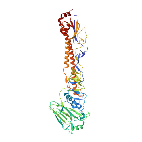 | |
UniProt | |||||
Find proteins for H9XCU1 (Influenza A virus) Explore H9XCU1 Go to UniProtKB: H9XCU1 | |||||
Entity Groups | |||||
| Sequence Clusters | 30% Identity50% Identity70% Identity90% Identity95% Identity100% Identity | ||||
| UniProt Group | H9XCU1 | ||||
Sequence AnnotationsExpand | |||||
| |||||
Entity ID: 2 | |||||
|---|---|---|---|---|---|
| Molecule | Chains | Length | 2D Diagram | Glycosylation | 3D Interactions |
| beta-D-mannopyranose-(1-4)-2-acetamido-2-deoxy-beta-D-glucopyranose-(1-4)-2-acetamido-2-deoxy-beta-D-glucopyranose | G, H, I, J, K G, H, I, J, K, L | 3 |  | N-Glycosylation | |
Glycosylation Resources | |||||
GlyTouCan: G15407YE GlyCosmos: G15407YE GlyGen: G15407YE | |||||
| Ligands 1 Unique | |||||
|---|---|---|---|---|---|
| ID | Chains | Name / Formula / InChI Key | 2D Diagram | 3D Interactions | |
| NAG (Subject of Investigation/LOI) Query on NAG | AA [auth D] BA [auth D] CA [auth E] DA [auth E] EA [auth E] | 2-acetamido-2-deoxy-beta-D-glucopyranose C8 H15 N O6 OVRNDRQMDRJTHS-FMDGEEDCSA-N |  | ||
| Length ( Å ) | Angle ( ˚ ) |
|---|---|
| a = 211.94 | α = 90 |
| b = 149.94 | β = 135.01 |
| c = 150.09 | γ = 90 |
| Software Name | Purpose |
|---|---|
| PHENIX | refinement |
| XDS | data reduction |
| Coot | model building |
| PHASER | phasing |
| Coot | model building |
RCSB PDB (citation) is hosted by
RCSB PDB is a member of the

