6OVO

Crystal structure of the unliganded PG10 TCR


Experimental Data Snapshot

  • Method: X-RAY DIFFRACTION
  • Resolution: 2.49 Å
  • R-Value Free: 0.222 
  • R-Value Work: 0.177 
  • R-Value Observed: 0.179 

wwPDB Validation   3D Report Full Report


This is version 1.2 of the entry. See complete history


Literature

A TCR beta-Chain Motif Biases toward Recognition of Human CD1 Proteins.

Reinink, P.Shahine, A.Gras, S.Cheng, T.Y.Farquhar, R.Lopez, K.Suliman, S.A.Reijneveld, J.F.Le Nours, J.Tan, L.L.Leon, S.R.Jimenez, J.Calderon, R.Lecca, L.Murray, M.B.Rossjohn, J.Moody, D.B.Van Rhijn, I.

(2019) J Immunol 203: 3395-3406

  • DOI: https://doi.org/10.4049/jimmunol.1900872
  • Primary Citation of Related Structures:  
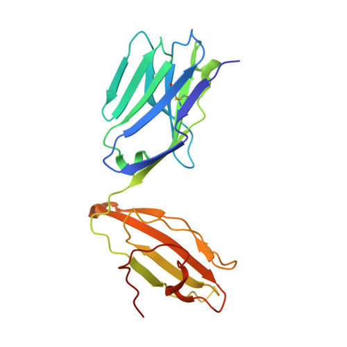
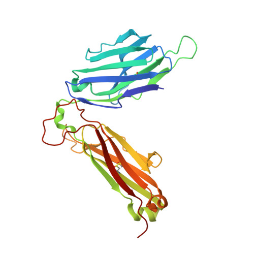
    6OVN, 6OVO

  • PubMed Abstract: 

    High-throughput TCR sequencing allows interrogation of the human TCR repertoire, potentially connecting TCR sequences to antigenic targets. Unlike the highly polymorphic MHC proteins, monomorphic Ag-presenting molecules such as MR1, CD1d, and CD1b present Ags to T cells with species-wide TCR motifs. CD1b tetramer studies and a survey of the 27 published CD1b-restricted TCRs demonstrated a TCR motif in humans defined by the TCR β-chain variable gene 4-1 (TRBV4-1) region. Unexpectedly, TRBV4-1 was involved in recognition of CD1b regardless of the chemical class of the carried lipid. Crystal structures of two CD1b-specific TRBV4-1 + TCRs show that germline-encoded residues in CDR1 and CDR3 regions of TRBV4-1-encoded sequences interact with each other and consolidate the surface of the TCR. Mutational studies identified a key positively charged residue in TRBV4-1 and a key negatively charged residue in CD1b that is shared with CD1c, which is also recognized by TRBV4-1 TCRs. These data show that one TCR V region can mediate a mechanism of recognition of two related monomorphic Ag-presenting molecules that does not rely on a defined lipid Ag.


  • Organizational Affiliation

    Department of Infectious Diseases and Immunology, Faculty of Veterinary Medicine, Utrecht University, 3584CL Utrecht, the Netherlands.


Macromolecules
Find similar proteins by:  (by identity cutoff)  |  3D Structure
Entity ID: 1
MoleculeChains Sequence LengthOrganismDetailsImage
Alpha Chain T-Cell Receptor PG10206Homo sapiensMutation(s): 0 
Gene Names: TRAV (TCRA)
Entity Groups  
Sequence Clusters30% Identity50% Identity70% Identity90% Identity95% Identity100% Identity
Sequence Annotations
Expand
  • Reference Sequence
Find similar proteins by:  (by identity cutoff)  |  3D Structure
Entity ID: 2
MoleculeChains Sequence LengthOrganismDetailsImage
Beta Chain T-Cell Receptor PG10247Homo sapiensMutation(s): 0 
Gene Names: TRBV (TCRB)
Entity Groups  
Sequence Clusters30% Identity50% Identity70% Identity90% Identity95% Identity100% Identity
Sequence Annotations
Expand
  • Reference Sequence
Experimental Data & Validation

Experimental Data

  • Method: X-RAY DIFFRACTION
  • Resolution: 2.49 Å
  • R-Value Free: 0.222 
  • R-Value Work: 0.177 
  • R-Value Observed: 0.179 
  • Space Group: P 61
Unit Cell:
Length ( Å )Angle ( ˚ )
a = 149.62α = 90
b = 149.62β = 90
c = 55.991γ = 120
Software Package:
Software NamePurpose
PHENIXrefinement
PDB_EXTRACTdata extraction
iMOSFLMdata reduction
Aimlessdata scaling
PHASERphasing

Structure Validation

View Full Validation Report


Entry History 

Deposition Data

Revision History  (Full details and data files)

  • Version 1.0: 2019-11-20
    Type: Initial release
  • Version 1.1: 2019-12-25
    Changes: Database references
  • Version 1.2: 2023-10-11
    Changes: Data collection, Database references, Refinement description