Highly Drug-Resistant HIV-1 Protease Mutant PRS17 Shows Enhanced Binding to Substrate Analogues.
Agniswamy, J., Kneller, D.W., Brothers, R., Wang, Y.F., Harrison, R.W., Weber, I.T.(2019) ACS Omega 4: 8707-8719
- PubMed: 31172041
- DOI: https://doi.org/10.1021/acsomega.9b00683
- Primary Citation of Related Structures:
6O48, 6O54, 6O57, 6O5A, 6O5X - PubMed Abstract:
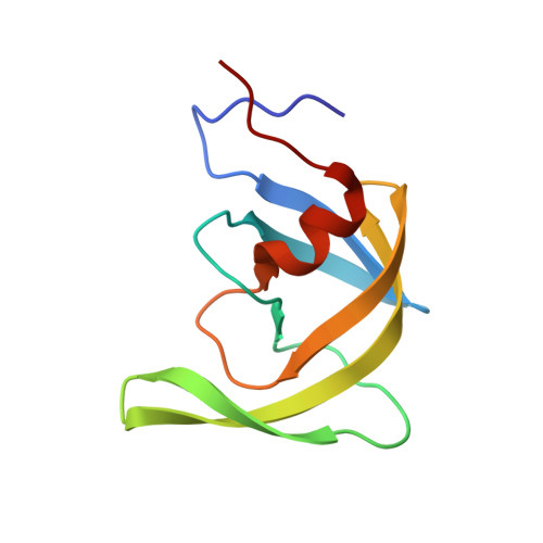
We report the structural analysis of highly drug-resistant human immunodeficiency virus protease (PR) variant PR S17 , rationally selected by machine learning, in complex with substrate analogues. Crystal structures were solved of inhibitor-free inactive PR S17 -D25N, wild-type PR/CA-p2 complex, and PR S17 in complex with substrate analogues, CA-p2 and p2-NC. Peptide analogues p2-NC and CA-p2 exhibit inhibition constants of 514 and 22 nM, respectively, for PR S17 or approximately 3-fold better than for PR. CA-p2 is a better inhibitor of PR S17 than are clinical inhibitors ( K i = 50-8390 nM) except for amprenavir ( K i = 11 nM). G48V resistance mutation induces curled flap tips in PR S17 -D25N structure. The inner P2-P2' residues of substrate analogues in PR S17 complexes maintain similar conformations to those of wild-type complex, while significant conformational changes are observed in the peripheral residues P3, P4' of CA-p2 and P3, P4, and P3' of p2-NC. The loss of β-branched side chain by V82S mutation initiates a shift in 80's loop and reshapes the S3/S3' subsite, which enhances substrate binding with new hydrogen bonds and van der Waals interactions that are absent in the wild-type structures. The steric hindrance caused by G48V mutation in the flap of PR S17 contributes to altered binding interactions of P3 Arg, P4' norleucine of CA-p2, and P4 and P3' of p2-NC with the addition of new hydrogen bonds and van der Waals contacts. The enhanced interaction of PR S17 with substrate analogues agrees with their relative inhibition, suggesting that this mutant improves substrate binding while decreasing affinity for clinical inhibitors.
Organizational Affiliation:
Department of Biology, Georgia State University, P.O. Box 4010, Atlanta, Georgia 30302, United States.