6NHL

Crystal structure of QueE from Escherichia coli


Experimental Data Snapshot

  • Method: X-RAY DIFFRACTION
  • Resolution: 2.10 Å
  • R-Value Free: 0.226 
  • R-Value Work: 0.173 
  • R-Value Observed: 0.176 

wwPDB Validation   3D Report Full Report


This is version 1.4 of the entry. See complete history


Literature

Crystal structure of AdoMet radical enzyme 7-carboxy-7-deazaguanine synthase from Escherichia coli suggests how modifications near [4Fe-4S] cluster engender flavodoxin specificity.

Grell, T.A.J.Bell, B.N.Nguyen, C.Dowling, D.P.Bruender, N.A.Bandarian, V.Drennan, C.L.

(2019) Protein Sci 28: 202-215

  • DOI: https://doi.org/10.1002/pro.3529
  • Primary Citation of Related Structures:  
    6NHL

  • PubMed Abstract: 

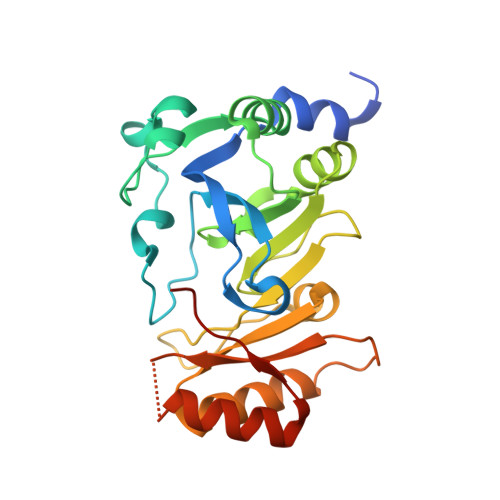
    7-Carboxy-7-deazaguanine synthase, QueE, catalyzes the radical mediated ring contraction of 6-carboxy-5,6,7,8-tetrahydropterin, forming the characteristic pyrrolopyrimidine core of all 7-deazaguanine natural products. QueE is a member of the S-adenosyl-L-methionine (AdoMet) radical enzyme superfamily, which harnesses the reactivity of radical intermediates to perform challenging chemical reactions. Members of the AdoMet radical enzyme superfamily utilize a canonical binding motif, a CX 3 CXϕC motif, to bind a [4Fe-4S] cluster, and a partial (β/α) 6 TIM barrel fold for the arrangement of AdoMet and substrates for catalysis. Although variations to both the cluster-binding motif and the core fold have been observed, visualization of drastic variations in the structure of QueE from Burkholderia multivorans called into question whether a re-haul of the defining characteristics of this superfamily was in order. Surprisingly, the structure of QueE from Bacillus subtilis revealed an architecture more reminiscent of the classical AdoMet radical enzyme. With these two QueE structures revealing varying degrees of alterations to the classical AdoMet fold, a new question arises: what is the purpose of these alterations? Here, we present the structure of a third QueE enzyme from Escherichia coli, which establishes the middle range of the spectrum of variation observed in these homologs. With these three homologs, we compare and contrast the structural architecture and make hypotheses about the role of these structural variations in binding and recognizing the biological reductant, flavodoxin. Broader impact statement: We know more about how enzymes are tailored for catalytic activity than about how enzymes are tailored to react with a physiological reductant. Here, we consider structural differences between three 7-carboxy-7-deazaguanine synthases and how these differences may be related to the interaction between these enzymes and their biological reductant, flavodoxin.


  • Organizational Affiliation

    Department of Chemistry, Massachusetts Institute of Technology, Cambridge, Massachusetts, 02139.


Macromolecules
Find similar proteins by:  (by identity cutoff)  |  3D Structure
Entity ID: 1
MoleculeChains Sequence LengthOrganismDetailsImage
7-carboxy-7-deazaguanine synthase
A, B
243Escherichia coli K-12Mutation(s): 0 
Gene Names: queEEC1303_c28920
EC: 4.3.99.3
Entity Groups  
Sequence Clusters30% Identity50% Identity70% Identity90% Identity95% Identity100% Identity
Sequence Annotations
Expand
  • Reference Sequence
Small Molecules
Ligands 5 Unique
IDChains Name / Formula / InChI Key2D Diagram3D Interactions
SF4
Query on SF4

Download Ideal Coordinates CCD File 
D [auth A],
G [auth B]
IRON/SULFUR CLUSTER
Fe4 S4
LJBDFODJNLIPKO-UHFFFAOYSA-N
PEG
Query on PEG

Download Ideal Coordinates CCD File 
E [auth A]DI(HYDROXYETHYL)ETHER
C4 H10 O3
MTHSVFCYNBDYFN-UHFFFAOYSA-N
GOL
Query on GOL

Download Ideal Coordinates CCD File 
H [auth B]GLYCEROL
C3 H8 O3
PEDCQBHIVMGVHV-UHFFFAOYSA-N
FE
Query on FE

Download Ideal Coordinates CCD File 
C [auth A]FE (III) ION
Fe
VTLYFUHAOXGGBS-UHFFFAOYSA-N
MG
Query on MG

Download Ideal Coordinates CCD File 
F [auth B]MAGNESIUM ION
Mg
JLVVSXFLKOJNIY-UHFFFAOYSA-N
Experimental Data & Validation

Experimental Data

  • Method: X-RAY DIFFRACTION
  • Resolution: 2.10 Å
  • R-Value Free: 0.226 
  • R-Value Work: 0.173 
  • R-Value Observed: 0.176 
  • Space Group: P 21 21 21
Unit Cell:
Length ( Å )Angle ( ˚ )
a = 70.264α = 90
b = 74.493β = 90
c = 103.714γ = 90
Software Package:
Software NamePurpose
PHENIXrefinement
HKL-2000data scaling
HKL-2000data reduction
HKL2Mapphasing
AutoSolphasing

Structure Validation

View Full Validation Report


Entry History & Funding Information

Deposition Data


Funding OrganizationLocationGrant Number
National Science Foundation (NSF, United States)United States1122374
National Institutes of Health/National Human Genome Research Institute (NIH/NHGRI)United StatesR35 GM126982 R01 GM72623 R35 GM126956 and R35 GM126982

Revision History  (Full details and data files)

  • Version 1.0: 2019-01-16
    Type: Initial release
  • Version 1.1: 2019-02-06
    Changes: Data collection, Refinement description
  • Version 1.2: 2019-05-08
    Changes: Data collection, Source and taxonomy
  • Version 1.3: 2019-11-27
    Changes: Author supporting evidence
  • Version 1.4: 2024-03-13
    Changes: Data collection, Database references, Derived calculations