6NAJ

Integrin AlphaVBeta3 ectodomain bound to Hr10 variant of the 10th domain of Fibronectin.


Experimental Data Snapshot

  • Method: X-RAY DIFFRACTION
  • Resolution: 3.10 Å
  • R-Value Free: 0.268 
  • R-Value Work: 0.238 
  • R-Value Observed: 0.239 

wwPDB Validation   3D Report Full Report


Ligand Structure Quality Assessment 


This is version 3.2 of the entry. See complete history


Literature

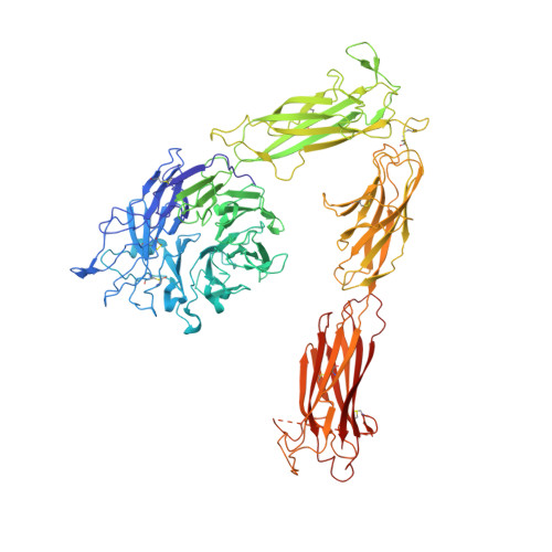
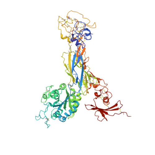
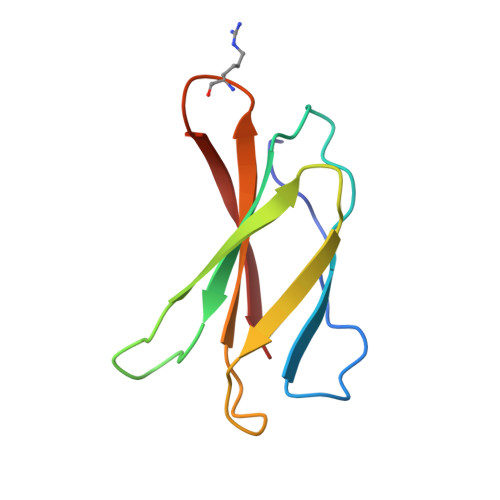
Structure-guided design of pure orthosteric inhibitors of alpha IIb beta 3 that prevent thrombosis but preserve hemostasis.

Adair, B.D.Alonso, J.L.van Agthoven, J.Hayes, V.Ahn, H.S.Yu, I.S.Lin, S.W.Xiong, J.P.Poncz, M.Arnaout, M.A.

(2020) Nat Commun 11: 398-398

  • DOI: https://doi.org/10.1038/s41467-019-13928-2
  • Primary Citation of Related Structures:  
    6NAJ

  • PubMed Abstract: 

    A prevailing dogma is that inhibition of vascular thrombosis by antagonizing platelet integrin αIIbβ3 cannot be achieved without compromising hemostasis, thus causing serious bleeding and increased morbidity and mortality. It is speculated that these adverse outcomes result from drug-induced activating conformational changes in αIIbβ3 but direct proof is lacking. Here, we report the structure-guided design of peptide Hr10 and a modified form of the partial agonist drug tirofiban that act as "pure" antagonists of αIIbβ3, i.e., they no longer induce the conformational changes in αIIbβ3. Both agents inhibit human platelet aggregation but preserve clot retraction. Hr10 and modified tirofiban are as effective as partial agonist drugs in inhibiting vascular thrombosis in humanized mice, but neither causes serious bleeding, establishing a causal link between partial agonism and impaired hemostasis. Pure orthosteric inhibitors of αIIbβ3 may thus provide safer alternatives for human therapy, and valuable tools to probe structure-activity relationships in integrins.


  • Organizational Affiliation

    Leukocyte Biology & Inflammation Program, and Structural Biology Program, Massachusetts General Hospital, Boston, MA, 02114, USA.


Macromolecules
Find similar proteins by:  (by identity cutoff)  |  3D Structure
Entity ID: 1
MoleculeChains Sequence LengthOrganismDetailsImage
Integrin alpha-V954Homo sapiensMutation(s): 0 
Gene Names: ITGAVMSK8VNRAVTNR
UniProt & NIH Common Fund Data Resources
Find proteins for P06756 (Homo sapiens)
Explore P06756 
Go to UniProtKB:  P06756
PHAROS:  P06756
GTEx:  ENSG00000138448 
Entity Groups  
Sequence Clusters30% Identity50% Identity70% Identity90% Identity95% Identity100% Identity
UniProt GroupP06756
Sequence Annotations
Expand
  • Reference Sequence
Find similar proteins by:  (by identity cutoff)  |  3D Structure
Entity ID: 2
MoleculeChains Sequence LengthOrganismDetailsImage
Integrin beta-3690Homo sapiensMutation(s): 0 
Gene Names: ITGB3GP3A
UniProt & NIH Common Fund Data Resources
Find proteins for P05106 (Homo sapiens)
Explore P05106 
Go to UniProtKB:  P05106
PHAROS:  P05106
GTEx:  ENSG00000259207 
Entity Groups  
Sequence Clusters30% Identity50% Identity70% Identity90% Identity95% Identity100% Identity
UniProt GroupP05106
Sequence Annotations
Expand
  • Reference Sequence
Find similar proteins by:  (by identity cutoff)  |  3D Structure
Entity ID: 3
MoleculeChains Sequence LengthOrganismDetailsImage
Fibronectin, HR10 variant90Homo sapiensMutation(s): 0 
Gene Names: FN1FN
UniProt & NIH Common Fund Data Resources
Find proteins for P02751 (Homo sapiens)
Explore P02751 
Go to UniProtKB:  P02751
PHAROS:  P02751
GTEx:  ENSG00000115414 
Entity Groups  
Sequence Clusters30% Identity50% Identity70% Identity90% Identity95% Identity100% Identity
UniProt GroupP02751
Sequence Annotations
Expand
  • Reference Sequence
Oligosaccharides

Help

Entity ID: 4
MoleculeChains Length2D Diagram Glycosylation3D Interactions
2-acetamido-2-deoxy-beta-D-glucopyranose-(1-4)-2-acetamido-2-deoxy-beta-D-glucopyranose
D, F, G, H, J
2N-Glycosylation
Glycosylation Resources
GlyTouCan:  G42666HT
GlyCosmos:  G42666HT
GlyGen:  G42666HT
Entity ID: 5
MoleculeChains Length2D Diagram Glycosylation3D Interactions
alpha-D-mannopyranose-(1-4)-beta-D-mannopyranose-(1-6)-[alpha-D-mannopyranose-(1-3)]beta-D-mannopyranose-(1-4)-2-acetamido-2-deoxy-beta-D-glucopyranose-(1-4)-2-acetamido-2-deoxy-beta-D-glucopyranose
E
6N-Glycosylation
Glycosylation Resources
GlyTouCan:  G93290PO
GlyCosmos:  G93290PO
GlyGen:  G93290PO
Entity ID: 6
MoleculeChains Length2D Diagram Glycosylation3D Interactions
beta-D-mannopyranose-(1-4)-2-acetamido-2-deoxy-beta-D-glucopyranose-(1-4)-2-acetamido-2-deoxy-beta-D-glucopyranose
I
3N-Glycosylation
Glycosylation Resources
GlyTouCan:  G15407YE
GlyCosmos:  G15407YE
GlyGen:  G15407YE
Small Molecules
Ligands 2 Unique
IDChains Name / Formula / InChI Key2D Diagram3D Interactions
NAG
Query on NAG

Download Ideal Coordinates CCD File 
K [auth A]
L [auth A]
M [auth A]
N [auth A]
O [auth A]
K [auth A],
L [auth A],
M [auth A],
N [auth A],
O [auth A],
U [auth B],
V [auth B]
2-acetamido-2-deoxy-beta-D-glucopyranose
C8 H15 N O6
OVRNDRQMDRJTHS-FMDGEEDCSA-N
MN
Query on MN

Download Ideal Coordinates CCD File 
P [auth A]
Q [auth A]
R [auth A]
S [auth A]
T [auth A]
P [auth A],
Q [auth A],
R [auth A],
S [auth A],
T [auth A],
W [auth B],
X [auth B],
Y [auth B]
MANGANESE (II) ION
Mn
WAEMQWOKJMHJLA-UHFFFAOYSA-N
Modified Residues  1 Unique
IDChains TypeFormula2D DiagramParent
HRG
Query on HRG
C
L-PEPTIDE LINKINGC7 H16 N4 O2ARG
Experimental Data & Validation

Experimental Data

  • Method: X-RAY DIFFRACTION
  • Resolution: 3.10 Å
  • R-Value Free: 0.268 
  • R-Value Work: 0.238 
  • R-Value Observed: 0.239 
  • Space Group: P 32 2 1
Unit Cell:
Length ( Å )Angle ( ˚ )
a = 129.713α = 90
b = 129.713β = 90
c = 308.198γ = 120
Software Package:
Software NamePurpose
PHENIXrefinement
PDB_EXTRACTdata extraction
HKL-2000data reduction
HKL-2000data scaling
PHENIXphasing

Structure Validation

View Full Validation Report


Ligand Structure Quality Assessment 


Entry History & Funding Information

Deposition Data


Funding OrganizationLocationGrant Number
National Institutes of Health/National Human Genome Research Institute (NIH/NHGRI)United StatesK01DK101628-04

Revision History  (Full details and data files)

  • Version 1.0: 2019-12-11
    Type: Initial release
  • Version 1.1: 2020-02-05
    Changes: Data collection, Database references
  • Version 2.0: 2020-07-29
    Type: Remediation
    Reason: Carbohydrate remediation
    Changes: Atomic model, Data collection, Derived calculations, Structure summary
  • Version 3.0: 2023-02-22
    Type: Coordinate replacement
    Reason: Ligand geometry
    Changes: Advisory, Atomic model, Data collection, Database references, Derived calculations, Refinement description, Source and taxonomy, Structure summary
  • Version 3.1: 2023-10-25
    Changes: Data collection, Refinement description
  • Version 3.2: 2023-11-15
    Changes: Data collection, Derived calculations