6N7U

apo-BDBV223 Fab


Experimental Data Snapshot

  • Method: X-RAY DIFFRACTION
  • Resolution: 2.03 Å
  • R-Value Free: 0.250 
  • R-Value Work: 0.235 
  • R-Value Observed: 0.235 

wwPDB Validation   3D Report Full Report

Currently 6N7U does not have a validation slider image.


This is version 1.3 of the entry. See complete history


Literature

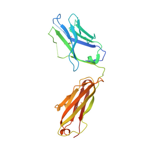
Cross-reactive neutralizing human survivor monoclonal antibody BDBV223 targets the ebolavirus stalk.

King, L.B.West, B.R.Moyer, C.L.Gilchuk, P.Flyak, A.Ilinykh, P.A.Bombardi, R.Hui, S.Huang, K.Bukreyev, A.Crowe Jr., J.E.Saphire, E.O.

(2019) Nat Commun 10: 1788-1788

  • DOI: https://doi.org/10.1038/s41467-019-09732-7
  • Primary Citation of Related Structures:  
    6N7J, 6N7U

  • PubMed Abstract: 

    Three Ebolavirus genus viruses cause lethal disease and lack targeted therapeutics: Ebola virus, Sudan virus and Bundibugyo virus. Monoclonal antibody (mAb) cocktails against the surface glycoprotein (GP) present a potential therapeutic strategy. Here we report two crystal structures of the antibody BDBV223, alone and complexed with its GP2 stalk epitope, an interesting site for therapeutic/vaccine design due to its high sequence conservation among ebolaviruses. BDBV223, identified in a human survivor of Bundibugyo virus disease, neutralizes both Bundibugyo virus and Ebola virus, but not Sudan virus. Importantly, the structure suggests that BDBV223 binding interferes with both the trimeric bundle assembly of GP and the viral membrane by stabilizing a conformation in which the monomers are separated by GP lifting or bending. Targeted mutagenesis of BDBV223 to enhance SUDV GP recognition indicates that additional determinants of antibody binding likely lie outside the visualized interactions, and perhaps involve quaternary assembly or membrane-interacting regions.


  • Organizational Affiliation

    Department of Immunology and Microbiology, The Scripps Research Institute, La Jolla, CA, 92037, USA.


Macromolecules
Find similar proteins by:  (by identity cutoff)  |  3D Structure
Entity ID: 1
MoleculeChains Sequence LengthOrganismDetailsImage
BDBV223 antibody heavy chain230Homo sapiensMutation(s): 0 
Entity Groups  
Sequence Clusters30% Identity50% Identity70% Identity90% Identity95% Identity100% Identity
Sequence Annotations
Expand
  • Reference Sequence
Find similar proteins by:  (by identity cutoff)  |  3D Structure
Entity ID: 2
MoleculeChains Sequence LengthOrganismDetailsImage
BDBV223 antibody light chain215Homo sapiensMutation(s): 0 
Entity Groups  
Sequence Clusters30% Identity50% Identity70% Identity90% Identity95% Identity100% Identity
Sequence Annotations
Expand
  • Reference Sequence
Experimental Data & Validation

Experimental Data

  • Method: X-RAY DIFFRACTION
  • Resolution: 2.03 Å
  • R-Value Free: 0.250 
  • R-Value Work: 0.235 
  • R-Value Observed: 0.235 
  • Space Group: P 21 21 21
Unit Cell:
Length ( Å )Angle ( ˚ )
a = 64.975α = 90
b = 75.678β = 90
c = 114.411γ = 90
Software Package:
Software NamePurpose
PHENIXrefinement
XDSdata reduction
XDSdata scaling
PHENIXphasing

Structure Validation

View Full Validation Report

Currently 6N7U does not have a validation slider image.


Entry History & Funding Information

Deposition Data


Funding OrganizationLocationGrant Number
National Institutes of Health/National Institute Of Allergy and Infectious Diseases (NIH/NIAID)United StatesU19 109762
National Institutes of Health/National Institute Of Allergy and Infectious Diseases (NIH/NIAID)United StatesU19 AI109711

Revision History  (Full details and data files)

  • Version 1.0: 2019-03-20
    Type: Initial release
  • Version 1.1: 2019-10-02
    Changes: Data collection, Database references
  • Version 1.2: 2019-12-18
    Changes: Author supporting evidence
  • Version 1.3: 2023-10-11
    Changes: Data collection, Database references, Refinement description