


Crystal structure of a Methionine aminopeptidase MetAP from Acinetobacter baumannii
Edwards, T.E., Mayclin, S.J., Lorimer, D.D., Horanyi, P.S., Seattle Structural Genomics Center for Infectious DiseaseTo be published.
Experimental Data Snapshot
wwPDB Validation 3D Report Full Report
Currently 6MRF does not have a validation slider image.
Entity ID: 1 | |||||
|---|---|---|---|---|---|
| Molecule | Chains | Sequence Length | Organism | Details | Image |
| Methionine aminopeptidase | 270 | Acinetobacter baumannii | Mutation(s): 0 Gene Names: map_1, map, map1, map_2, A7M79_08180, A7M90_04815, A7N09_12540, ABUW_1234, APD06_11055, AZE33_14285... EC: 3.4.11.18 | 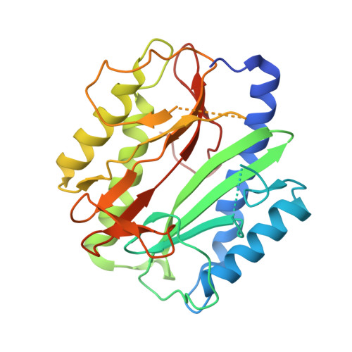 | |
UniProt | |||||
Find proteins for V5VCW7 (Acinetobacter baumannii) Explore V5VCW7 Go to UniProtKB: V5VCW7 | |||||
Entity Groups | |||||
| Sequence Clusters | 30% Identity50% Identity70% Identity90% Identity95% Identity100% Identity | ||||
| UniProt Group | V5VCW7 | ||||
Sequence AnnotationsExpand | |||||
| |||||
| Ligands 2 Unique | |||||
|---|---|---|---|---|---|
| ID | Chains | Name / Formula / InChI Key | 2D Diagram | 3D Interactions | |
| EDO Query on EDO | B [auth A], C [auth A] | 1,2-ETHANEDIOL C2 H6 O2 LYCAIKOWRPUZTN-UHFFFAOYSA-N |  | ||
| NA Query on NA | D [auth A] | SODIUM ION Na FKNQFGJONOIPTF-UHFFFAOYSA-N |  | ||
| Length ( Å ) | Angle ( ˚ ) |
|---|---|
| a = 52.77 | α = 90 |
| b = 53.69 | β = 90 |
| c = 84.27 | γ = 90 |
| Software Name | Purpose |
|---|---|
| XDS | data reduction |
| XSCALE | data scaling |
| PHASER | phasing |
| PHENIX | refinement |
| PDB_EXTRACT | data extraction |
Currently 6MRF does not have a validation slider image.
RCSB PDB (citation) is hosted by
RCSB PDB is a member of the

