6ML8

Crystal structure of hemagglutinin from H1N1 Influenza A virus A/Denver/57 bound to the C05 antibody


Experimental Data Snapshot

  • Method: X-RAY DIFFRACTION
  • Resolution: 2.92 Å
  • R-Value Free: 0.264 
  • R-Value Work: 0.201 
  • R-Value Observed: 0.204 

wwPDB Validation   3D Report Full Report


This is version 2.2 of the entry. See complete history


Literature

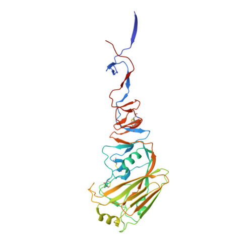
Structural characterisation of hemagglutinin from seven Influenza A H1N1 strains reveal diversity in the C05 antibody recognition site.

Ghafoori, S.M.Petersen, G.F.Conrady, D.G.Calhoun, B.M.Stigliano, M.Z.Z.Baydo, R.O.Grice, R.Abendroth, J.Lorimer, D.D.Edwards, T.E.Forwood, J.K.

(2023) Sci Rep 13: 6940-6940

  • DOI: https://doi.org/10.1038/s41598-023-33529-w
  • Primary Citation of Related Structures:  
    6D8W, 6ML8, 6MYA, 6N41, 6ONA, 6OSR, 7JP4, 7JPD

  • PubMed Abstract: 

    Influenza virus (IV) causes several outbreaks of the flu each year resulting in an economic burden to the healthcare system in the billions of dollars. Several influenza pandemics have occurred during the last century and estimated to have caused 100 million deaths. There are four genera of IV, A (IVA), B (IVB), C (IVC), and D (IVD), with IVA being the most virulent to the human population. Hemagglutinin (HA) is an IVA surface protein that allows the virus to attach to host cell receptors and enter the cell. Here we have characterised the high-resolution structures of seven IVA HAs, with one in complex with the anti-influenza head-binding antibody C05. Our analysis revealed conserved receptor binding residues in all structures, as seen in previously characterised IV HAs. Amino acid conservation is more prevalent on the stalk than the receptor binding domain (RBD; also called the head domain), allowing the virus to escape from antibodies targeting the RBD. The equivalent site of C05 antibody binding to A/Denver/57 HA appears hypervariable in the other H1N1 IV HAs. Modifications within this region appear to disrupt binding of the C05 antibody, as these HAs no longer bind the C05 antibody by analytical SEC. Our study brings new insights into the structural and functional recognition of IV HA proteins and can contribute to further development of anti-influenza vaccines.


  • Organizational Affiliation

    School of Dentistry and Medical Sciences, Charles Sturt University, Wagga Wagga, NSW, 2650, Australia.


Macromolecules
Find similar proteins by:  (by identity cutoff)  |  3D Structure
Entity ID: 1
MoleculeChains Sequence LengthOrganismDetailsImage
Hemagglutinin330Influenza A virus (A/Denver/1957(H1N1))Mutation(s): 0 
Gene Names: HA
UniProt
Find proteins for Q2IBI1 (Influenza A virus)
Explore Q2IBI1 
Go to UniProtKB:  Q2IBI1
Entity Groups  
Sequence Clusters30% Identity50% Identity70% Identity90% Identity95% Identity100% Identity
UniProt GroupQ2IBI1
Sequence Annotations
Expand
  • Reference Sequence
Find similar proteins by:  (by identity cutoff)  |  3D Structure
Entity ID: 2
MoleculeChains Sequence LengthOrganismDetailsImage
Hemagglutinin165Influenza A virus (A/Denver/1957(H1N1))Mutation(s): 0 
Gene Names: HA
UniProt
Find proteins for Q2IBI1 (Influenza A virus)
Explore Q2IBI1 
Go to UniProtKB:  Q2IBI1
Entity Groups  
Sequence Clusters30% Identity50% Identity70% Identity90% Identity95% Identity100% Identity
UniProt GroupQ2IBI1
Sequence Annotations
Expand
  • Reference Sequence
Find similar proteins by:  (by identity cutoff)  |  3D Structure
Entity ID: 3
MoleculeChains Sequence LengthOrganismDetailsImage
C05 antibody Fab heavy chainC [auth H]247Homo sapiensMutation(s): 0 
Entity Groups  
Sequence Clusters30% Identity50% Identity70% Identity90% Identity95% Identity100% Identity
Sequence Annotations
Expand
  • Reference Sequence
Find similar proteins by:  (by identity cutoff)  |  3D Structure
Entity ID: 4
MoleculeChains Sequence LengthOrganismDetailsImage
C05 antibody Fab light chainD [auth L]214Homo sapiensMutation(s): 0 
Entity Groups  
Sequence Clusters30% Identity50% Identity70% Identity90% Identity95% Identity100% Identity
Sequence Annotations
Expand
  • Reference Sequence
Oligosaccharides

Help

Entity ID: 5
MoleculeChains Length2D Diagram Glycosylation3D Interactions
alpha-D-mannopyranose-(1-3)-beta-D-mannopyranose-(1-4)-2-acetamido-2-deoxy-beta-D-glucopyranose-(1-4)-2-acetamido-2-deoxy-beta-D-glucopyranoseE [auth C]4N-Glycosylation
Glycosylation Resources
GlyTouCan:  G81315DD
GlyCosmos:  G81315DD
GlyGen:  G81315DD
Entity ID: 6
MoleculeChains Length2D Diagram Glycosylation3D Interactions
beta-D-mannopyranose-(1-4)-2-acetamido-2-deoxy-beta-D-glucopyranose-(1-4)-2-acetamido-2-deoxy-beta-D-glucopyranoseF [auth D]3N-Glycosylation
Glycosylation Resources
GlyTouCan:  G15407YE
GlyCosmos:  G15407YE
GlyGen:  G15407YE
Entity ID: 7
MoleculeChains Length2D Diagram Glycosylation3D Interactions
2-acetamido-2-deoxy-beta-D-glucopyranose-(1-4)-2-acetamido-2-deoxy-beta-D-glucopyranoseG [auth E]2N-Glycosylation
Glycosylation Resources
GlyTouCan:  G42666HT
GlyCosmos:  G42666HT
GlyGen:  G42666HT
Experimental Data & Validation

Experimental Data

Unit Cell:
Length ( Å )Angle ( ˚ )
a = 118.46α = 90
b = 118.46β = 90
c = 162.44γ = 120
Software Package:
Software NamePurpose
XDSdata reduction
XSCALEdata scaling
MOLREPphasing
PHENIXrefinement
PDB_EXTRACTdata extraction

Structure Validation

View Full Validation Report


Entry History 

Deposition Data

Revision History  (Full details and data files)

  • Version 1.0: 2018-10-17
    Type: Initial release
  • Version 2.0: 2020-07-29
    Type: Remediation
    Reason: Carbohydrate remediation
    Changes: Atomic model, Data collection, Derived calculations, Structure summary
  • Version 2.1: 2023-05-24
    Changes: Database references, Derived calculations, Structure summary
  • Version 2.2: 2023-10-25
    Changes: Data collection, Refinement description