Halogen Bond Interactions of Novel HIV-1 Protease Inhibitors (PI) (GRL-001-15 and GRL-003-15) with the Flap of Protease Are Critical for Their Potent Activity against Wild-Type HIV-1 and Multi-PI-Resistant Variants.
Hattori, S.I., Hayashi, H., Bulut, H., Rao, K.V., Nyalapatla, P.R., Hasegawa, K., Aoki, M., Ghosh, A.K., Mitsuya, H.(2019) Antimicrob Agents Chemother 63
- PubMed: 30962341
- DOI: https://doi.org/10.1128/AAC.02635-18
- Primary Citation of Related Structures:
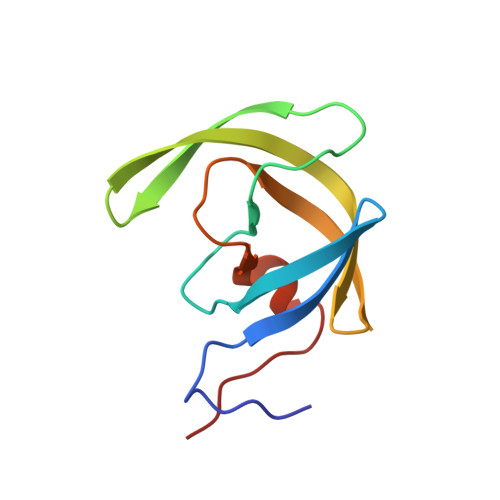
6MCR, 6MCS - PubMed Abstract:
We generated two novel nonpeptidic HIV-1 protease inhibitors (PIs), GRL-001-15 and GRL-003-15, which contain unique crown-like tetrahydropyranofuran (Crn-THF) and P2'-cyclopropyl-aminobenzothiazole (Cp-Abt) moieties as P2 and P2' ligands, respectively. GRL-001-15 and GRL-003-15 have meta -monofluorophenyl and para -monofluorophenyl at the P1 site, respectively, exert highly potent activity against wild-type HIV-1 with 50% effective concentrations (EC 50 s) of 57 and 50 pM, respectively, and have favorable cytotoxicity profiles with 50% cytotoxic concentrations (CC 50 s) of 38 and 11 μM, respectively. The activity of GRL-001-15 against multi-PI-resistant HIV-1 variants was generally greater than that of GRL-003-15. The EC 50 of GRL-001-15 against an HIV-1 variant that was highly resistant to multiple PIs, including darunavir (DRV) (HIV-1 DRV R P30 ), was 0.17 nM, and that of GRL-003-15 was 3.3 nM, while DRV was much less active, with an EC 50 of 216 nM. The emergence of HIV-1 variants resistant to GRL-001-15 and GRL-003-15 was significantly delayed compared to that of variants resistant to selected PIs, including DRV. Structural analyses of wild-type protease (PR WT ) complexed with the novel PIs revealed that GRL-001-15's meta -fluorine atom forms halogen bond interactions (2.9 and 3.0 Å) with Gly49 and Ile50, respectively, of the protease flap region and with Pro81' (2.7 and 3.2 Å), which is located close to the protease active site, and that two fluorine atoms of GRL-142-13 form multiple halogen bond interactions with Gly49, Ile50, Pro81', Ile82', and Arg8'. In contrast, GRL-003-15 forms halogen bond interactions with Pro81' alone, suggesting that the reduced antiviral activity of GRL-003-15 is due to the loss of the interactions with the flap region.
Organizational Affiliation:
Department of Refractory Viral Infections, National Center for Global Health and Medicine Research Institute, Tokyo, Japan.