6LT6

Crystal structure of rhesus macaque MHC class I molecule Mamu-B*05104 complexed with lysophosphatidylcholine


Experimental Data Snapshot

  • Method: X-RAY DIFFRACTION
  • Resolution: 2.15 Å
  • R-Value Free: 0.232 
  • R-Value Work: 0.182 
  • R-Value Observed: 0.184 

wwPDB Validation   3D Report Full Report


Ligand Structure Quality Assessment 


This is version 1.2 of the entry. See complete history


Literature

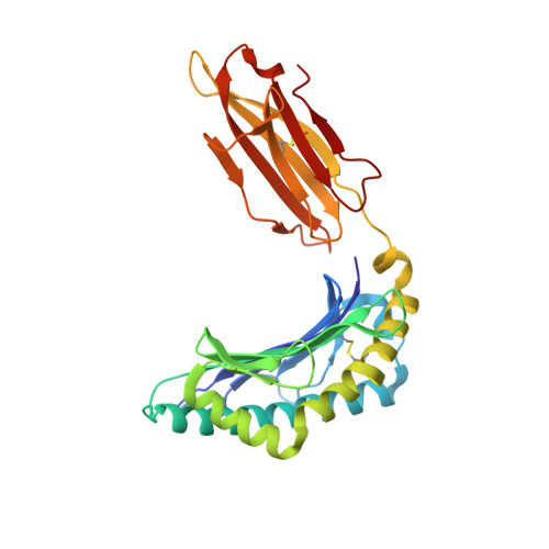
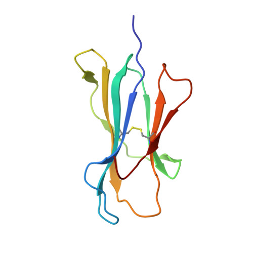
Crystal structures of lysophospholipid-bound MHC class I molecules.

Shima, Y.Morita, D.Mizutani, T.Mori, N.Mikami, B.Sugita, M.

(2020) J Biol Chem 295: 6983-6991

  • DOI: https://doi.org/10.1074/jbc.RA119.011932
  • Primary Citation of Related Structures:  
    6LAH, 6LAM, 6LB2, 6LT6

  • PubMed Abstract: 

    Newly synthesized major histocompatibility complex (MHC) class I proteins are stabilized in the endoplasmic reticulum (ER) by binding 8-10-mer-long self-peptide antigens that are provided by transporter associated with antigen processing (TAP). These MHC class I:peptide complexes then exit the ER and reach the plasma membrane, serving to sustain the steady-state MHC class I expression on the cell surface. A novel subset of MHC class I molecules that preferentially bind lipid-containing ligands rather than conventional peptides was recently identified. The primate classical MHC class I allomorphs, Mamu-B*098 and Mamu-B*05104, are capable of binding the N -myristoylated 5-mer (C14-Gly-Gly-Ala-Ile-Ser) or 4-mer (C14-Gly-Gly-Ala-Ile) lipopeptides derived from the N -myristoylated SIV Nef protein, respectively, and of activating lipopeptide antigen-specific cytotoxic T lymphocytes. We herein demonstrate that Mamu-B*098 samples lysophosphatidylethanolamine and lysophosphatidylcholine containing up to a C20 fatty acid in the ER. The X-ray crystal structures of Mamu-B*098 and Mamu-B*05104 complexed with lysophospholipids at high resolution revealed that the B and D pockets in the antigen-binding grooves of these MHC class I molecules accommodate these lipids through a monoacylglycerol moiety. Consistent with the capacity to bind cellular lipid ligands, these two MHC class I molecules did not require TAP function for cell-surface expression. Collectively, these results indicate that peptide- and lipopeptide-presenting MHC class I subsets use distinct sources of endogenous ligands.


  • Organizational Affiliation

    Laboratory of Cell Regulation, Institute for Frontier Life and Medical Sciences, Kyoto University, 53 Kawahara-cho, Shogoin, Sakyo-ku, Kyoto 606-8507, Japan.


Macromolecules
Find similar proteins by:  (by identity cutoff)  |  3D Structure
Entity ID: 1
MoleculeChains Sequence LengthOrganismDetailsImage
MHC class I antigen276Macaca mulattaMutation(s): 2 
Gene Names: Mamu-BB
UniProt
Find proteins for B2ZHY7 (Macaca mulatta)
Explore B2ZHY7 
Go to UniProtKB:  B2ZHY7
Entity Groups  
Sequence Clusters30% Identity50% Identity70% Identity90% Identity95% Identity100% Identity
UniProt GroupB2ZHY7
Sequence Annotations
Expand
  • Reference Sequence
Find similar proteins by:  (by identity cutoff)  |  3D Structure
Entity ID: 2
MoleculeChains Sequence LengthOrganismDetailsImage
Beta-2-microglobulin100Macaca mulattaMutation(s): 0 
Gene Names: B2M
UniProt
Find proteins for Q6V7J5 (Macaca mulatta)
Explore Q6V7J5 
Go to UniProtKB:  Q6V7J5
Entity Groups  
Sequence Clusters30% Identity50% Identity70% Identity90% Identity95% Identity100% Identity
UniProt GroupQ6V7J5
Sequence Annotations
Expand
  • Reference Sequence
Small Molecules
Ligands 3 Unique
IDChains Name / Formula / InChI Key2D Diagram3D Interactions
EKG (Subject of Investigation/LOI)
Query on EKG

Download Ideal Coordinates CCD File 
C [auth A](2R)-2,3-dihydroxypropyl hexadecanoate
C19 H38 O4
QHZLMUACJMDIAE-GOSISDBHSA-N
EDO
Query on EDO

Download Ideal Coordinates CCD File 
D [auth A]
E [auth A]
F [auth A]
G [auth B]
H [auth B]
D [auth A],
E [auth A],
F [auth A],
G [auth B],
H [auth B],
I [auth B],
J [auth B],
K [auth B]
1,2-ETHANEDIOL
C2 H6 O2
LYCAIKOWRPUZTN-UHFFFAOYSA-N
NA
Query on NA

Download Ideal Coordinates CCD File 
L [auth B]SODIUM ION
Na
FKNQFGJONOIPTF-UHFFFAOYSA-N
Experimental Data & Validation

Experimental Data

  • Method: X-RAY DIFFRACTION
  • Resolution: 2.15 Å
  • R-Value Free: 0.232 
  • R-Value Work: 0.182 
  • R-Value Observed: 0.184 
  • Space Group: P 21 21 21
Unit Cell:
Length ( Å )Angle ( ˚ )
a = 54.451α = 90
b = 81.028β = 90
c = 106.108γ = 90
Software Package:
Software NamePurpose
PHENIXrefinement
HKL-2000data reduction
HKL-2000data scaling
MOLREPphasing

Structure Validation

View Full Validation Report


Ligand Structure Quality Assessment 


Entry History & Funding Information

Deposition Data


Funding OrganizationLocationGrant Number
Japan Society for the Promotion of Science (JSPS)Japan18K07172
Japan Society for the Promotion of Science (JSPS)Japan17H05791
Japan Society for the Promotion of Science (JSPS)Japan18K19563
Japan Society for the Promotion of Science (JSPS)Japan18H02852
Japan Society for the Promotion of Science (JSPS)Japan19H04805

Revision History  (Full details and data files)

  • Version 1.0: 2020-04-22
    Type: Initial release
  • Version 1.1: 2020-05-27
    Changes: Database references
  • Version 1.2: 2023-11-29
    Changes: Data collection, Database references, Refinement description