6LRA

The complex structure of PHF core domain peptide of tau and antibody's Fab domain.


Experimental Data Snapshot

  • Method: X-RAY DIFFRACTION
  • Resolution: 1.90 Å
  • R-Value Free: 0.249 
  • R-Value Work: 0.188 
  • R-Value Observed: 0.191 

wwPDB Validation   3D Report Full Report


This is version 1.1 of the entry. See complete history


Literature

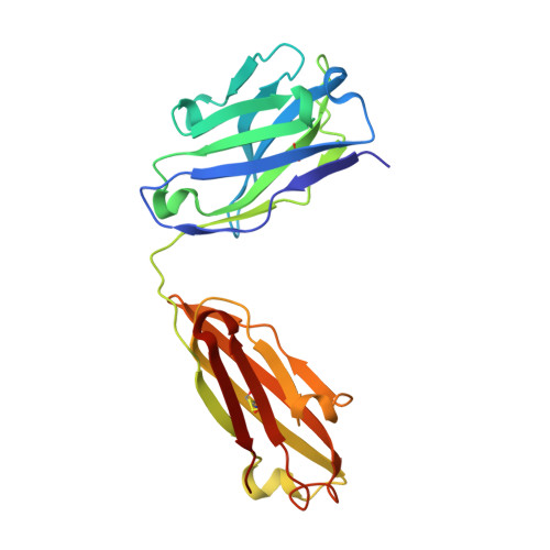
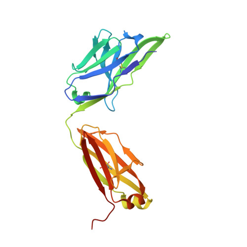
Crystal structure of the human tau PHF core domain VQIINK complexed with the Fab domain of monoclonal antibody Tau2r3.

Tsuchida, T.Susa, K.Kibiki, T.Tsuchiya, T.Miyamoto, K.In, Y.Minoura, K.Taniguchi, T.Ishida, T.Tomoo, K.

(2020) FEBS Lett 

  • DOI: https://doi.org/10.1002/1873-3468.13791
  • Primary Citation of Related Structures:  
    6LRA

  • PubMed Abstract: 

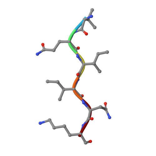
    Neurofibrillary tangles formed by abnormally aggregated tau protein are a histopathological feature of tauopathies. A tau aggregation inhibitor is a potential therapeutic agent for tauopathies. In this study, we prepared a monoclonal antibody for tau, monoclonal antibody to tau protein (Tau2r3), using as epitope the 272 GGKVQIINKKLD 283 peptide in the microtubule-binding domain of tau, the key region mediating tau aggregation. We show that Tau2r3 clearly inhibits tau aggregation. To analyze the inhibition mechanism of Tau2r3, we solved the crystal structure of the Fab domain of Tau2r3 (Fab2r3) in complex with the VQIINK peptide. In the Fab2r3-VQIINK structure, the second and sixth polar residues and the fourth hydrophobic residue of VQIINK are crucial for binding to Fab2r3. The structural data for the Fab2r3-VQIINK complex could contribute to the design of new tau aggregation inhibitors.


  • Organizational Affiliation

    Department of Physical Chemistry, Osaka University of Pharmaceutical Sciences, Takatsuki, Japan.


Macromolecules
Find similar proteins by:  (by identity cutoff)  |  3D Structure
Entity ID: 1
MoleculeChains Sequence LengthOrganismDetailsImage
Fab Heavy ChainA [auth H]218Mus musculusMutation(s): 0 
Entity Groups  
Sequence Clusters30% Identity50% Identity70% Identity90% Identity95% Identity100% Identity
Sequence Annotations
Expand
  • Reference Sequence
Find similar proteins by:  (by identity cutoff)  |  3D Structure
Entity ID: 2
MoleculeChains Sequence LengthOrganismDetailsImage
Fab Light ChainB [auth L]220Mus musculusMutation(s): 0 
Entity Groups  
Sequence Clusters30% Identity50% Identity70% Identity90% Identity95% Identity100% Identity
Sequence Annotations
Expand
  • Reference Sequence

Find similar proteins by:  Sequence   |   3D Structure  

Entity ID: 3
MoleculeChains Sequence LengthOrganismDetailsImage
VQIINK6Homo sapiensMutation(s): 0 
UniProt & NIH Common Fund Data Resources
Find proteins for P10636 (Homo sapiens)
Explore P10636 
Go to UniProtKB:  P10636
PHAROS:  P10636
GTEx:  ENSG00000186868 
Entity Groups  
UniProt GroupP10636
Sequence Annotations
Expand
  • Reference Sequence
Experimental Data & Validation

Experimental Data

  • Method: X-RAY DIFFRACTION
  • Resolution: 1.90 Å
  • R-Value Free: 0.249 
  • R-Value Work: 0.188 
  • R-Value Observed: 0.191 
  • Space Group: P 21 21 21
Unit Cell:
Length ( Å )Angle ( ˚ )
a = 59.65α = 90
b = 70.68β = 90
c = 118.37γ = 90
Software Package:
Software NamePurpose
REFMACrefinement
PDB_EXTRACTdata extraction
Cootmodel building
MOLREPphasing
CrystalCleardata reduction
CrystalCleardata scaling

Structure Validation

View Full Validation Report


Entry History 

Deposition Data

Revision History  (Full details and data files)

  • Version 1.0: 2020-08-26
    Type: Initial release
  • Version 1.1: 2023-11-29
    Changes: Data collection, Database references, Refinement description