6LKS

Effects of zinc ion on oligomerization and pH stability of influenza virus hemagglutinin


Experimental Data Snapshot

  • Method: X-RAY DIFFRACTION
  • Resolution: 3.24 Å
  • R-Value Free: 0.295 
  • R-Value Work: 0.265 
  • R-Value Observed: 0.266 

wwPDB Validation   3D Report Full Report


Ligand Structure Quality Assessment 


This is version 1.2 of the entry. See complete history


Literature

Divalent cation-induced conformational changes of influenza virus hemagglutinin.

Seok, J.H.Kim, H.Lee, D.B.An, J.S.Kim, E.J.Lee, J.H.Chung, M.S.Kim, K.H.

(2020) Sci Rep 10: 15457-15457

  • DOI: https://doi.org/10.1038/s41598-020-72368-x
  • Primary Citation of Related Structures:  
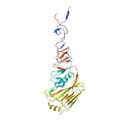
    6LKS

  • PubMed Abstract: 

    Divalent cations Cu 2+ and Zn 2+ can prevent the viral growth in mammalian cells during influenza infection, and viral titers decrease significantly on a copper surface. The underlying mechanisms include DNA damage by radicals, modulation of viral protease, M1 or neuraminidase, and morphological changes in viral particles. However, the molecular mechanisms underlying divalent cation-mediated antiviral activities are unclear. An unexpected observation of this study was that a Zn 2+ ion is bound by Glu68 and His137 residues at the head regions of two neighboring trimers in the crystal structure of hemagglutinin (HA) derived from A/Thailand/CU44/2006. The binding of Zn 2+ at high concentrations induced multimerization of HA and decreased its acid stability. The acid-induced conformational change of HA occurred even at neutral pH in the presence of Zn 2+ . The fusion of viral and host endosomal membranes requires substantial conformational changes in HA upon exposure to acidic pH. Therefore, our results suggest that binding of Zn 2+ may facilitate the conformational changes of HA, analogous to that induced by acidic pH.


  • Organizational Affiliation

    Department of Biotechnology and Bioinformatics, Korea University, Sejong, 30019, Korea.


Macromolecules
Find similar proteins by:  (by identity cutoff)  |  3D Structure
Entity ID: 1
MoleculeChains Sequence LengthOrganismDetailsImage
Hemagglutinin HA1 chain330Influenza A virus (A/Thailand/CU44/2006(H1N1))Mutation(s): 0 
Gene Names: HA
UniProt
Find proteins for A7LI25 (Influenza A virus)
Explore A7LI25 
Go to UniProtKB:  A7LI25
Entity Groups  
Sequence Clusters30% Identity50% Identity70% Identity90% Identity95% Identity100% Identity
UniProt GroupA7LI25
Sequence Annotations
Expand
  • Reference Sequence
Find similar proteins by:  (by identity cutoff)  |  3D Structure
Entity ID: 2
MoleculeChains Sequence LengthOrganismDetailsImage
Hemagglutinin HA2 chain183Influenza A virus (A/Thailand/CU44/2006(H1N1))Mutation(s): 0 
Gene Names: HA
UniProt
Find proteins for A7LI25 (Influenza A virus)
Explore A7LI25 
Go to UniProtKB:  A7LI25
Entity Groups  
Sequence Clusters30% Identity50% Identity70% Identity90% Identity95% Identity100% Identity
UniProt GroupA7LI25
Sequence Annotations
Expand
  • Reference Sequence
Oligosaccharides

Help

Entity ID: 3
MoleculeChains Length2D Diagram Glycosylation3D Interactions
2-acetamido-2-deoxy-beta-D-glucopyranose-(1-2)-alpha-D-mannopyranose-(1-3)-[alpha-D-mannopyranose-(1-6)]beta-D-mannopyranose-(1-4)-2-acetamido-2-deoxy-beta-D-glucopyranose-(1-4)-2-acetamido-2-deoxy-beta-D-glucopyranose
M, S, V
6N-Glycosylation
Glycosylation Resources
GlyTouCan:  G61751GZ
GlyCosmos:  G61751GZ
GlyGen:  G61751GZ
Entity ID: 4
MoleculeChains Length2D Diagram Glycosylation3D Interactions
2-acetamido-2-deoxy-beta-D-glucopyranose-(1-2)-alpha-D-mannopyranose-(1-3)-beta-D-mannopyranose-(1-4)-2-acetamido-2-deoxy-beta-D-glucopyranose-(1-4)-2-acetamido-2-deoxy-beta-D-glucopyranose
N, P
5N-Glycosylation
Glycosylation Resources
GlyTouCan:  G10133VD
GlyCosmos:  G10133VD
Entity ID: 5
MoleculeChains Length2D Diagram Glycosylation3D Interactions
alpha-D-mannopyranose-(1-3)-beta-D-mannopyranose-(1-4)-2-acetamido-2-deoxy-beta-D-glucopyranose-(1-4)-2-acetamido-2-deoxy-beta-D-glucopyranose
O, Q, R, T, U
O, Q, R, T, U, X
4N-Glycosylation
Glycosylation Resources
GlyTouCan:  G81315DD
GlyCosmos:  G81315DD
GlyGen:  G81315DD
Entity ID: 6
MoleculeChains Length2D Diagram Glycosylation3D Interactions
beta-D-mannopyranose-(1-4)-2-acetamido-2-deoxy-beta-D-glucopyranose-(1-4)-2-acetamido-2-deoxy-beta-D-glucopyranose
W
3N-Glycosylation
Glycosylation Resources
GlyTouCan:  G15407YE
GlyCosmos:  G15407YE
GlyGen:  G15407YE
Small Molecules
Ligands 2 Unique
IDChains Name / Formula / InChI Key2D Diagram3D Interactions
NAG (Subject of Investigation/LOI)
Query on NAG

Download Ideal Coordinates CCD File 
AA [auth K]
CA [auth A]
DA [auth A]
EA [auth A]
FA [auth A]
AA [auth K],
CA [auth A],
DA [auth A],
EA [auth A],
FA [auth A],
GA [auth A],
HA [auth C],
IA [auth E],
JA [auth E],
KA [auth E],
LA [auth E],
MA [auth G],
NA [auth G],
OA [auth G],
QA [auth I],
RA [auth I],
SA [auth I],
Y [auth K],
Z [auth K]
2-acetamido-2-deoxy-beta-D-glucopyranose
C8 H15 N O6
OVRNDRQMDRJTHS-FMDGEEDCSA-N
ZN (Subject of Investigation/LOI)
Query on ZN

Download Ideal Coordinates CCD File 
BA [auth K],
PA [auth G],
TA [auth I]
ZINC ION
Zn
PTFCDOFLOPIGGS-UHFFFAOYSA-N
Experimental Data & Validation

Experimental Data

  • Method: X-RAY DIFFRACTION
  • Resolution: 3.24 Å
  • R-Value Free: 0.295 
  • R-Value Work: 0.265 
  • R-Value Observed: 0.266 
  • Space Group: C 1 2 1
Unit Cell:
Length ( Å )Angle ( ˚ )
a = 215.71α = 90
b = 124.27β = 102.909
c = 214.53γ = 90
Software Package:
Software NamePurpose
ADSCdata collection
PHENIXrefinement
XDSdata reduction
XDSdata scaling
PHENIXphasing

Structure Validation

View Full Validation Report


Ligand Structure Quality Assessment 


Entry History & Funding Information

Deposition Data


Funding OrganizationLocationGrant Number
National Research Foundation (NRF, Korea)Korea, Republic Of2010-0029242

Revision History  (Full details and data files)

  • Version 1.0: 2020-10-21
    Type: Initial release
  • Version 1.1: 2020-10-28
    Changes: Database references
  • Version 1.2: 2023-11-22
    Changes: Data collection, Database references, Refinement description