6LKG

two-component system protein mediate signal transduction


Experimental Data Snapshot

  • Method: X-RAY DIFFRACTION
  • Resolution: 1.95 Å
  • R-Value Free: 0.225 
  • R-Value Work: 0.204 
  • R-Value Observed: 0.205 

wwPDB Validation   3D Report Full Report


Ligand Structure Quality Assessment 


This is version 1.2 of the entry. See complete history


Literature

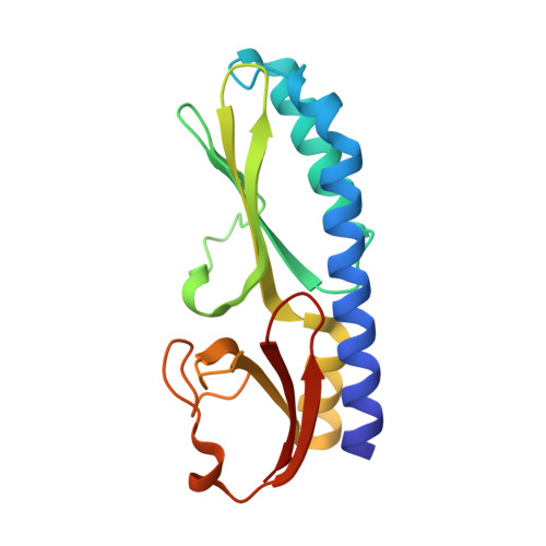
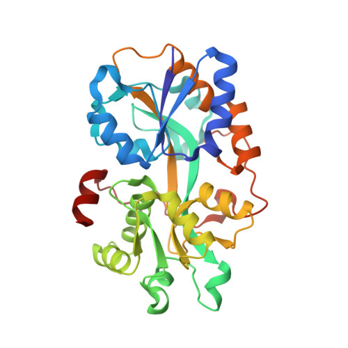
Interface switch mediates signal transmission in a two-component system.

Wang, M.Guo, Q.Zhu, K.Fang, B.Yang, Y.Teng, M.Li, X.Tao, Y.

(2020) Proc Natl Acad Sci U S A 117: 30433-30440

  • DOI: https://doi.org/10.1073/pnas.1912080117
  • Primary Citation of Related Structures:  
    6LKG, 6LKH, 6LKI, 6LKJ, 6LKK, 6LKL

  • PubMed Abstract: 

    Two-component systems (TCS), which typically consist of a membrane-embedded histidine kinase and a cytoplasmic response regulator, are the dominant signaling proteins for transduction of environmental stimuli into cellular response pathways in prokaryotic cells. HptRSA is a recently identified TCS consisting of the G6P-associated sensor protein (HptA), transmembrane histidine kinase (HptS), and cytoplasmic effector (HptR). HptRSA mediates glucose-6-phosphate (G6P) uptake to support Staphylococcus aureus growth and multiplication within various host cells. How the mechanism by which HptRSA perceives G6P and triggers a downstream response has remained elusive. Here, we solved the HptA structures in apo and G6P-bound states. G6P binding in the cleft between two HptA domains caused a conformational closing movement. The solved structures of HptA in complex with the periplasmic domain of HptS showed that HptA interacts with HptS through both constitutive and switchable interfaces. The G6P-free form of HptA binds to the membrane-distal side of the HptS periplasmic domain (HptSp), resulting in a parallel conformation of the HptSp protomer pair. However, once HptA associates with G6P, its intramolecular domain closure switches the HptA-HptSp contact region into the membrane-proximal domain, which causes rotation and closure of the C termini of each HptSp protomer. Through biochemical and growth assays of HptA and HptS mutant variants, we proposed a distinct mechanism of interface switch-mediated signaling transduction. Our results provide mechanistic insights into bacterial nutrient sensing and expand our understanding of the activation modes by which TCS communicates external signals.


  • Organizational Affiliation

    Ministry of Education Key Laboratory for Membraneless Organelles & Cellular Dynamics, Hefei National Laboratory for Physical Sciences at the Microscale, School of Life Sciences, Division of Life Sciences and Medicine, University of Science and Technology of China, 230027 Hefei, P.R. China.


Macromolecules
Find similar proteins by:  (by identity cutoff)  |  3D Structure
Entity ID: 1
MoleculeChains Sequence LengthOrganismDetailsImage
Sensor protein kinase HptSA [auth B],
D
172Staphylococcus aureus subsp. aureus NCTC 8325Mutation(s): 0 
Gene Names: hptSSAOUHSC_00185
EC: 2.7.13.3
UniProt
Find proteins for Q2G1E0 (Staphylococcus aureus (strain NCTC 8325 / PS 47))
Explore Q2G1E0 
Go to UniProtKB:  Q2G1E0
Entity Groups  
Sequence Clusters30% Identity50% Identity70% Identity90% Identity95% Identity100% Identity
UniProt GroupQ2G1E0
Sequence Annotations
Expand
  • Reference Sequence
Find similar proteins by:  (by identity cutoff)  |  3D Structure
Entity ID: 2
MoleculeChains Sequence LengthOrganismDetailsImage
ABC transporter, solute-binding proteinB [auth A],
C
294Staphylococcus aureusMutation(s): 0 
Gene Names: E4U00_07700EP54_06205EQ90_12025HMPREF3211_02751NCTC10654_00249NCTC10702_00414RK64_01575
UniProt
Find proteins for X5DVD1 (Staphylococcus aureus)
Explore X5DVD1 
Go to UniProtKB:  X5DVD1
Entity Groups  
Sequence Clusters30% Identity50% Identity70% Identity90% Identity95% Identity100% Identity
UniProt GroupX5DVD1
Sequence Annotations
Expand
  • Reference Sequence
Small Molecules
Experimental Data & Validation

Experimental Data

  • Method: X-RAY DIFFRACTION
  • Resolution: 1.95 Å
  • R-Value Free: 0.225 
  • R-Value Work: 0.204 
  • R-Value Observed: 0.205 
  • Space Group: P 1 21 1
Unit Cell:
Length ( Å )Angle ( ˚ )
a = 72.3α = 90
b = 89.97β = 100.87
c = 92.827γ = 90
Software Package:
Software NamePurpose
PHENIXrefinement
PDB_EXTRACTdata extraction
HKL-3000data reduction
HKL-3000data scaling
PHENIXphasing

Structure Validation

View Full Validation Report


Ligand Structure Quality Assessment 


Entry History 

Deposition Data

Revision History  (Full details and data files)

  • Version 1.0: 2020-12-02
    Type: Initial release
  • Version 1.1: 2020-12-16
    Changes: Database references
  • Version 1.2: 2024-03-27
    Changes: Data collection, Database references, Refinement description