6LBE

Crystal structure of bony fish MHC class I binding beta2M-2 for 2.6 angstrom


Experimental Data Snapshot

  • Method: X-RAY DIFFRACTION
  • Resolution: 2.60 Å
  • R-Value Free: 0.254 
  • R-Value Work: 0.169 
  • R-Value Observed: 0.173 

wwPDB Validation   3D Report Full Report


This is version 1.1 of the entry. See complete history


Literature

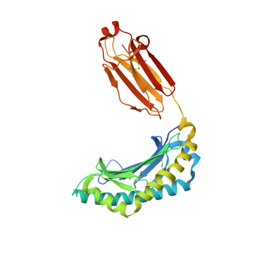
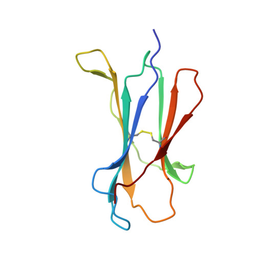
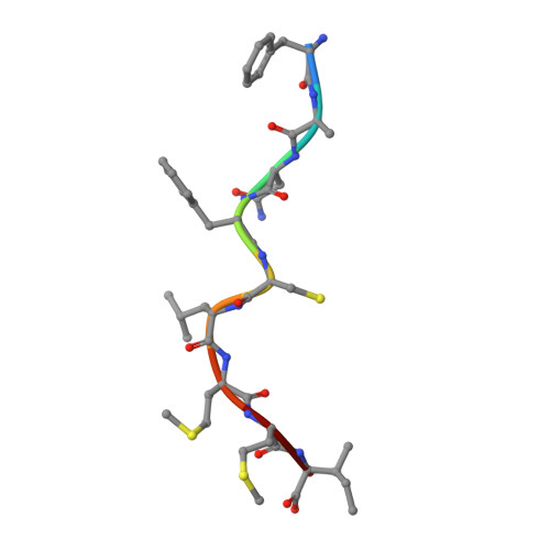
The Mechanism of beta 2m Molecule-Induced Changes in the Peptide Presentation Profile in a Bony Fish.

Li, Z.Zhang, N.Ma, L.Zhang, L.Meng, G.Xia, C.

(2020) iScience 23: 101119-101119

  • DOI: https://doi.org/10.1016/j.isci.2020.101119
  • Primary Citation of Related Structures:  
    5H5Z, 6LBE

  • PubMed Abstract: 

    Contemporary antigen presentation knowledge is based on the existence of a single β2m locus, and a classical MHC class I forms a complex with a peptide (i.e., pMHC-I) to trigger CTL immunity. However, two β2m loci have been found in diploid bony fish; the function of the two β2m molecules is unclear. Here, we determined the variant peptide profiles originating from different products of the β2m loci binding to the same MHC-I molecule and further solved the crystal structures of the two pMHC-I molecules (i.e., pCtid-UAA-β2m-2 and pCtid-UAA-β2m-1-II). Of note, in pCtid-UAA-β2m-2, a unique hydrogen bond network formed in the bottom of the peptide-binding groove (PBG) led to α2-helix drift, ultimately leading to structural changes in the PBG. The mechanism of the change in peptide presentation profiles by β2m molecules is illustrated. The results are also of great significance for antivirus and antitumor functions in cold-blooded vertebrates and even humans.


  • Organizational Affiliation

    Department of Microbiology and Immunology, College of Veterinary Medicine, China Agricultural University, Beijing, 100193, China.


Macromolecules
Find similar proteins by:  (by identity cutoff)  |  3D Structure
Entity ID: 1
MoleculeChains Sequence LengthOrganismDetailsImage
MHC class I antigen
A, C
275Ctenopharyngodon idellaMutation(s): 0 
Gene Names: UAA106
UniProt
Find proteins for Q65XY8 (Ctenopharyngodon idella)
Explore Q65XY8 
Go to UniProtKB:  Q65XY8
Entity Groups  
Sequence Clusters30% Identity50% Identity70% Identity90% Identity95% Identity100% Identity
UniProt GroupQ65XY8
Sequence Annotations
Expand
  • Reference Sequence
Find similar proteins by:  (by identity cutoff)  |  3D Structure
Entity ID: 2
MoleculeChains Sequence LengthOrganismDetailsImage
Beta-2-microglobulin
B, D
99Ctenopharyngodon idellaMutation(s): 0 
Gene Names: b2m-b
UniProt
Find proteins for A0A3G2VUI3 (Ctenopharyngodon idella)
Explore A0A3G2VUI3 
Go to UniProtKB:  A0A3G2VUI3
Entity Groups  
Sequence Clusters30% Identity50% Identity70% Identity90% Identity95% Identity100% Identity
UniProt GroupA0A3G2VUI3
Sequence Annotations
Expand
  • Reference Sequence

Find similar proteins by:  Sequence   |   3D Structure  

Entity ID: 3
MoleculeChains Sequence LengthOrganismDetailsImage
9-mer peptide from RNA-DIRECTED RNA POLYMERASE L
E, F
9synthetic constructMutation(s): 0 
UniProt
Find proteins for Q91DR9 (Spring viremia of carp virus)
Explore Q91DR9 
Go to UniProtKB:  Q91DR9
Entity Groups  
UniProt GroupQ91DR9
Sequence Annotations
Expand
  • Reference Sequence
Experimental Data & Validation

Experimental Data

  • Method: X-RAY DIFFRACTION
  • Resolution: 2.60 Å
  • R-Value Free: 0.254 
  • R-Value Work: 0.169 
  • R-Value Observed: 0.173 
  • Space Group: P 1
Unit Cell:
Length ( Å )Angle ( ˚ )
a = 53.19α = 64.94
b = 65.27β = 75.67
c = 69.71γ = 73.61
Software Package:
Software NamePurpose
PHENIXrefinement
iMOSFLMdata reduction
SCALAdata scaling
PHASERphasing

Structure Validation

View Full Validation Report


Entry History & Funding Information

Deposition Data


Funding OrganizationLocationGrant Number
National Natural Science Foundation of ChinaChinaGrant 31572653

Revision History  (Full details and data files)

  • Version 1.0: 2020-11-04
    Type: Initial release
  • Version 1.1: 2023-11-22
    Changes: Data collection, Database references, Refinement description