6LAW

MicroED structure of proteinase K at 1.50A determained using crystal lamellas prepared by focused ion beam milling

  • Classification: HYDROLASE
  • Organism(s): Parengyodontium album
  • Mutation(s): Yes 

  • Deposited: 2019-11-13 Released: 2019-12-04 
  • Deposition Author(s): Zhou, H., Luo, Z., Li, X.
  • Funding Organization(s): National Natural Science Foundation of China, Ministry of Science and Technology (China)

Experimental Data Snapshot

  • Method: ELECTRON CRYSTALLOGRAPHY
  • Resolution: 1.50 Å
  • R-Value Free: 0.225 
  • R-Value Work: 0.187 
  • R-Value Observed: 0.189 

wwPDB Validation   3D Report Full Report


This is version 1.0 of the entry. See complete history


Literature

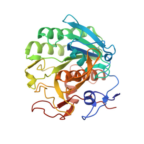
Using focus ion beam to prepare crystal lamella for electron diffraction.

Zhou, H.Luo, Z.Li, X.

(2019) J Struct Biol 205: 59-64

  • DOI: https://doi.org/10.1016/j.jsb.2019.02.004
  • Primary Citation of Related Structures:  
    6LAV, 6LAW

  • PubMed Abstract: 

    Electron diffraction provides a powerful tool to solve the structures of small protein crystals. However, strong interactions between the electrons and the materials limit the application of the electron crystallographic method on large protein crystals with micrometer or larger sizes. Here, we used the focused ion beam (FIB) equipped on the scanning electron microscope (SEM) to mill a large crystal to thin lamella. The influences of the milling on the crystal lamella were observed and investigated, including radiation damage on the crystal surface during the FIB imaging, deformation of the thin crystal lamella, and variation in the diffraction intensities under electron radiation. These observations provide important information to optimize the FIB milling, and hence is important to obtain high-quality crystal samples for routine structure determination of protein crystals using the electron cryo-microscope.


  • Organizational Affiliation

    Key Laboratory of Protein Sciences (Tsinghua University), Ministry of Education, Beijing, China; School of Life Sciences, Tsinghua University, Beijing, China.


Macromolecules
Find similar proteins by:  (by identity cutoff)  |  3D Structure
Entity ID: 1
MoleculeChains Sequence LengthOrganismDetailsImage
Proteinase K279Parengyodontium albumMutation(s): 1 
EC: 3.4.21.64
UniProt
Find proteins for P06873 (Parengyodontium album)
Explore P06873 
Go to UniProtKB:  P06873
Entity Groups  
Sequence Clusters30% Identity50% Identity70% Identity90% Identity95% Identity100% Identity
UniProt GroupP06873
Sequence Annotations
Expand
  • Reference Sequence
Small Molecules
Ligands 1 Unique
IDChains Name / Formula / InChI Key2D Diagram3D Interactions
SO4
Query on SO4

Download Ideal Coordinates CCD File 
B [auth A]SULFATE ION
O4 S
QAOWNCQODCNURD-UHFFFAOYSA-L
Experimental Data & Validation

Experimental Data

  • Method: ELECTRON CRYSTALLOGRAPHY
  • Resolution: 1.50 Å
  • R-Value Free: 0.225 
  • R-Value Work: 0.187 
  • R-Value Observed: 0.189 
  • Space Group: P 43 21 2
Unit Cell:
Length ( Å )Angle ( ˚ )
a = 69.314α = 90
b = 69.314β = 90
c = 104.116γ = 90
Software Package:
Software NamePurpose
REFMACrefinement
PDB_EXTRACTdata extraction

Structure Validation

View Full Validation Report


Entry History & Funding Information

Deposition Data


Funding OrganizationLocationGrant Number
National Natural Science Foundation of ChinaChina31570730
National Natural Science Foundation of ChinaChina31722015
Ministry of Science and Technology (China)China2016YFA0501102
Ministry of Science and Technology (China)China2016YFA0501902

Revision History  (Full details and data files)

  • Version 1.0: 2019-12-04
    Type: Initial release