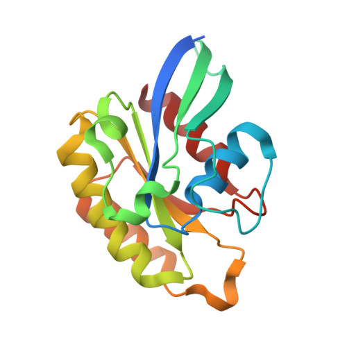
Crystal structure of the GDP-bound GTPase domain of Rab5a from Leishmania donovani.
Zohib, M., Maheshwari, D., Pal, R.K., Freitag-Pohl, S., Biswal, B.K., Pohl, E., Arora, A.(2020) Acta Crystallogr F Struct Biol Commun 76: 544-556
- PubMed: 33135673
- DOI: https://doi.org/10.1107/S2053230X20013722
- Primary Citation of Related Structures:
6L6O - PubMed Abstract:
Eukaryotic Rab5s are highly conserved small GTPase-family proteins that are involved in the regulation of early endocytosis. Leishmania donovani Rab5a regulates the sorting of early endosomes that are involved in the uptake of essential nutrients through fluid-phase endocytosis. Here, the 1.80 Å resolution crystal structure of the N-terminal GTPase domain of L. donovani Rab5a in complex with GDP is presented. The crystal structure determination was enabled by the design of specific single-site mutations and two deletions that were made to stabilize the protein for previous NMR studies. The structure of LdRab5a shows the canonical GTPase fold, with a six-stranded central mixed β-sheet surrounded by five α-helices. The positions of the Switch I and Switch II loops confirm an open conformation, as expected in the absence of the γ-phosphate. However, in comparison to other GTP-bound and GDP-bound homologous proteins, the Switch I region traces a unique disposition in LdRab5a. One magnesium ion is bound to the protein at the GTP-binding site. Molecular-dynamics simulations indicate that the GDP-bound structure exhibits higher stability than the apo structure. The GDP-bound LdRab5a structure presented here will aid in efforts to unravel its interactions with its regulators, including the guanine nucleotide-exchange factor, and will lay the foundation for a structure-based search for specific inhibitors.
Organizational Affiliation:
Molecular and Structural Biology Division, CSIR - Central Drug Research Institute, Lucknow 226 031, India.