


Crystal structure of E136F mutant of Xanthine-guanine phosphoribosyltransferase from Yersinia pestis
Lankipalli, S., Ramagopal, U.A.To be published.
Experimental Data Snapshot
wwPDB Validation 3D Report Full Report

Entity ID: 1 | |||||
|---|---|---|---|---|---|
| Molecule | Chains | Sequence Length | Organism | Details | Image |
| Xanthine phosphoribosyltransferase | 153 | Yersinia pestis | Mutation(s): 1 Gene Names: gpt, YPO3225, y0963, YP_0708 EC: 2.4.2.22 | 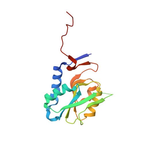 | |
UniProt | |||||
Find proteins for Q8ZC05 (Yersinia pestis) Explore Q8ZC05 Go to UniProtKB: Q8ZC05 | |||||
Entity Groups | |||||
| Sequence Clusters | 30% Identity50% Identity70% Identity90% Identity95% Identity100% Identity | ||||
| UniProt Group | Q8ZC05 | ||||
Sequence AnnotationsExpand | |||||
| |||||
| Ligands 2 Unique | |||||
|---|---|---|---|---|---|
| ID | Chains | Name / Formula / InChI Key | 2D Diagram | 3D Interactions | |
| GOL Query on GOL | F [auth B] | GLYCEROL C3 H8 O3 PEDCQBHIVMGVHV-UHFFFAOYSA-N |  | ||
| CL Query on CL | C [auth A], D [auth B], E [auth B] | CHLORIDE ION Cl VEXZGXHMUGYJMC-UHFFFAOYSA-M |  | ||
| Length ( Å ) | Angle ( ˚ ) |
|---|---|
| a = 56.38 | α = 90 |
| b = 96.65 | β = 90 |
| c = 51.029 | γ = 90 |
| Software Name | Purpose |
|---|---|
| Aimless | data scaling |
| REFMAC | refinement |
| PDB_EXTRACT | data extraction |
| MOSFLM | data reduction |
| MOLREP | phasing |
RCSB PDB (citation) is hosted by
RCSB PDB is a member of the

