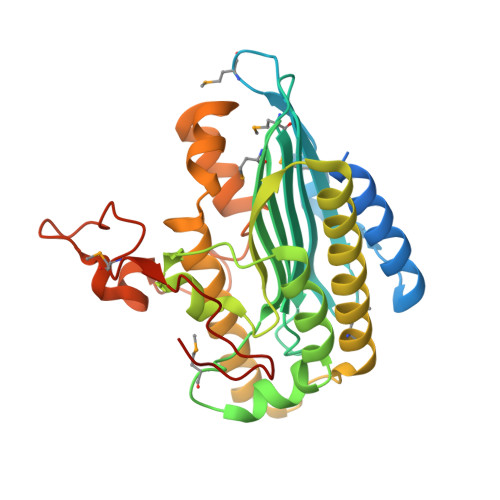
Crystal structure of phytochromobilin synthase in complex with biliverdin IX alpha , a key enzyme in the biosynthesis of phytochrome.
Sugishima, M., Wada, K., Fukuyama, K., Yamamoto, K.(2020) J Biol Chem 295: 771-782
- PubMed: 31822504
- DOI: https://doi.org/10.1074/jbc.RA119.011431
- Primary Citation of Related Structures:
6KMD, 6KME - PubMed Abstract:
Phytochromobilin (PΦB) is a red/far-red light sensory pigment in plant phytochrome. PΦB synthase is a ferredoxin-dependent bilin reductase (FDBR) that catalyzes the site-specific reduction of bilins, which are sensory and photosynthesis pigments, and produces PΦB from biliverdin, a heme-derived linear tetrapyrrole pigment. Here, we determined the crystal structure of tomato PΦB synthase in complex with biliverdin at 1.95 Å resolution. The overall structure of tomato PΦB synthase was similar to those of other FDBRs, except for the addition of a long C-terminal loop and short helices. The structure further revealed that the C-terminal loop is part of the biliverdin-binding pocket and that two basic residues in the C-terminal loop form salt bridges with the propionate groups of biliverdin. This suggested that the C-terminal loop is involved in the interaction with ferredoxin and biliverdin. The configuration of biliverdin bound to tomato PΦB synthase differed from that of biliverdin bound to other FDBRs, and its orientation in PΦB synthase was inverted relative to its orientation in the other FDBRs. Structural and enzymatic analyses disclosed that two aspartic acid residues, Asp-123 and Asp-263, form hydrogen bonds with water molecules and are essential for the site-specific A-ring reduction of biliverdin. On the basis of these observations and enzymatic assays with a V121A PΦB synthase variant, we propose the following mechanistic product release mechanism: PΦB synthase-catalyzed stereospecific reduction produces 2( R )-PΦB, which when bound to PΦB synthase collides with the side chain of Val-121, releasing 2( R )-PΦB from the synthase.
Organizational Affiliation:
Department of Medical Biochemistry, Kurume University School of Medicine, Kurume, Fukuoka 830-0011, Japan sugishima_masakazu@med.kurume-u.ac.jp.